
Sebaceous Gland Collection
The sebaceous gland, a tiny but crucial component of our skin, plays a significant role in both maintaining its health and causing various skin disorders


All Professionally Made to Order for Quick Shipping
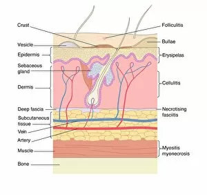
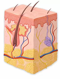
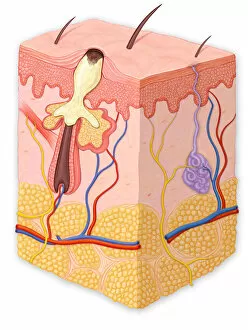
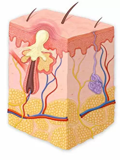
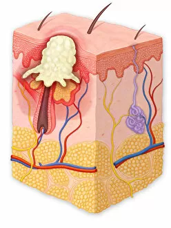
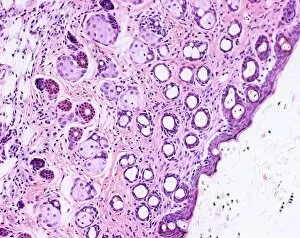
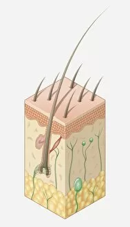
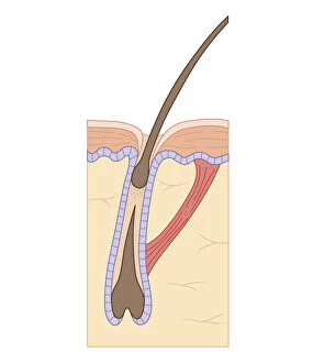
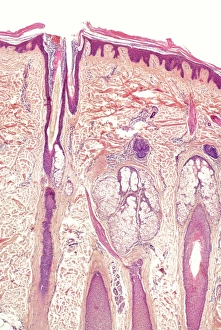
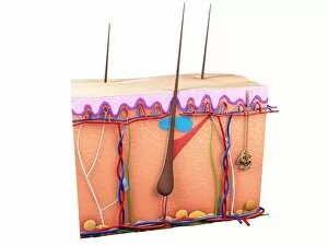
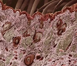
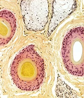
The sebaceous gland, a tiny but crucial component of our skin, plays a significant role in both maintaining its health and causing various skin disorders. A normal cross section of the skin reveals its intricate layers, with the sebaceous glands nestled within. These glands produce an oily substance called sebum that helps keep our skin moisturized and protected. However, when these glands become overactive or clogged, they can lead to pesky skin issues like blackheads, papules, pustules, and whiteheads. In a cross section showing a blackhead, we witness the accumulation of excess oil and dead skin cells within the pore. Similarly, a papule showcases inflammation caused by blocked sebaceous glands. A pustule exhibits an infected lesion filled with pus due to bacterial growth in the blocked gland. On the other hand, a whitehead occurs when trapped sebum remains beneath the surface of the skin. To better understand this complex system within our bodies, illustrations depicting the structure of human skin and hair prove invaluable. Light micrographs reveal intricate details of both our precious dermis and hair follicles while digital illustrations provide clear insights into their composition. Cross-section biomedical illustrations further enhance comprehension by showcasing detailed depictions of both healthy hair follicles during their growth phase as well as overall structures found in human skin. Exploring these captivating visuals not only deepens our knowledge about sebaceous glands but also highlights their significance in maintaining healthy-looking skin while shedding light on common dermatological concerns we may encounter along life's journey.