
Secretion Collection
"Unveiling the Mysteries of Secretion: From Brain to Ink and Beyond" Delving into the depths of our brain


All Professionally Made to Order for Quick Shipping
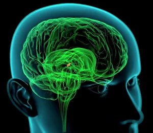
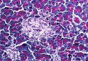
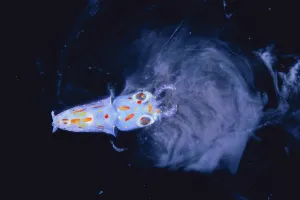
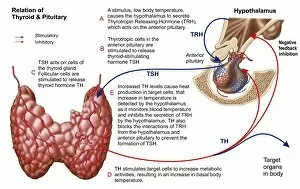
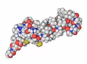
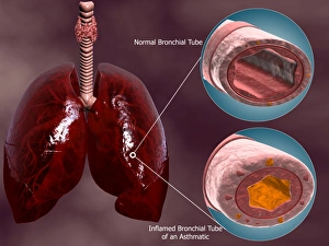
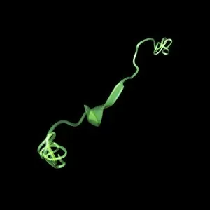
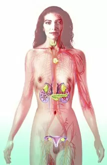
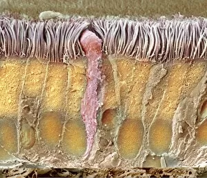
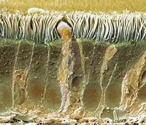
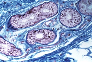
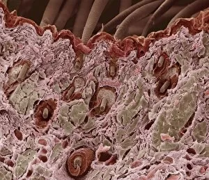
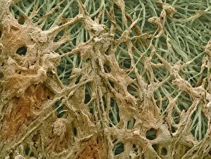
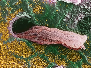
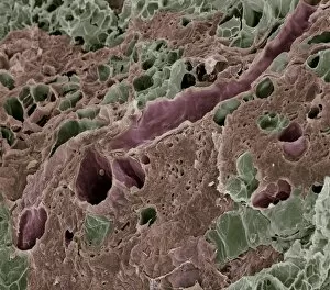
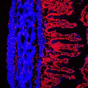
"Unveiling the Mysteries of Secretion: From Brain to Ink and Beyond" Delving into the depths of our brain, the medulla oblongata orchestrates a symphony of secretions that regulate vital bodily functions. Like an intricate artwork, the pancreatic islet of Langerhans reveals its hidden talent for secreting hormones essential for glucose metabolism. Three enigmatic adder stones hold ancient secrets, rumored to possess mystical powers related to secretion in folklore and mythology. In the depths of captivity, a tiny spear-like Bleekers Squid larva surprises us by secreting ink as a defense mechanism - nature's own disappearing act. Unraveling the complex relationship between the thyroid and pituitary gland unveils their crucial role in regulating hormone secretion throughout our body. Peering through electron microscopy lenses, we witness the delicate trachea lining at microscopic levels - a fascinating world where secretion takes place. With each SEM image revealing intricate details, we explore how normal and asthmatic bronchioles differ within cross-sections of our respiratory system's trachea. The parathyroid hormone molecule emerges as a key player in maintaining calcium balance through its precise secretion control mechanisms. Connecting back to their earlier mention, we revisit the intriguing relationship between thyroid and pituitary glands - two powerhouses driving hormonal secretions within us. A striking visual comparison showcases both healthy and asthmatic bronchioles alongside lungs within tracheal cross-sections - highlighting how secretion impacts respiratory health. In this captivating journey through various aspects of "secretion, " from brain function to squid ink defense mechanisms, we uncover remarkable connections across different realms of science and nature alike.