Fraud Blocker Skip to main content

Sense Collection (#7)

"Exploring the Multifaceted World of Sense

Background imageSense Collection: Human brain and senses, artwork

Human brain and senses, artwork
Human brain and senses. Artwork of a human brain, seen obliquely from the front, with the five senses shown. These are the visual organs (vision, the eyes), the olfactory organ (smell, the nose)

Background imageSense Collection: Senses within the brain, 16th century C017 / 6996

Senses within the brain, 16th century C017 / 6996
Senses within the brain. 16th-century woodcut of a mans head in profile, showing his brain, with various parts named, including memory, cognition, fantasia, sensus

Background imageSense Collection: Ampullary cupula, artwork

Ampullary cupula, artwork
Ampullary cupula. Computer artwork showing how the ampullary cupula in the human vestibular system of the inner ear senses head rotation (top) and forward motion (bottom)

Background imageSense Collection: Metarhodopsin molecule F006 / 9709

Metarhodopsin molecule F006 / 9709
Metarhodopsin II molecule, molecular model. This is one of the intermediary breakdown products formed when light falls on the photoreceptor pigment rhodopsin

Background imageSense Collection: Opsin molecule F006 / 9665

Opsin molecule F006 / 9665
Opsin. Molecular model of a ligand-free opsin molecule. Opsins are found in photoreceptor cells (rods and cones) in the retina of the eye

Background imageSense Collection: Rhodopsin molecule F006 / 9325

Rhodopsin molecule F006 / 9325
Rhodopsin molecule. Molecular model of the rhodopsin complex. Rhodopsin (also called visual purple) is a pigment found in the rod photoreceptor cells in the retina of the eye

Background imageSense Collection: Structure of the cochlea, artwork

Structure of the cochlea, artwork
Structure of the cochlea. Computer artwork of a section through the organ of Corti, the auditory sense organ that lines the spiral of the cochlea in the inner ear

Background imageSense Collection: Hearing centre, artwork

Hearing centre, artwork
Hearing centre. Computer artwork showing the area of the brains temporal lobe that is associated with hearing (red). This area receives auditory input from the cochlea (coiled)

Background imageSense Collection: Tropical spider detecting prey C016 / 8424

Tropical spider detecting prey C016 / 8424
Tropical spider detecting prey. Spider with its foot on a silk strand to sense prey in its web. Photographed in Napo Province, Amazon rainforest, Ecuador

Background imageSense Collection: Gall midge eye, SEM C016 / 9394

Gall midge eye, SEM C016 / 9394
Gall midge eye. Coloured scanning electron micrograph (SEM) of the compound eye of a gall midge (family Cecidomyiidae), showing the individual lenses (ommatidia)

Background imageSense Collection: Owl butterfly head, SEM C016 / 9432

Owl butterfly head, SEM C016 / 9432
Owl butterfly head. Coloured scanning electron micrograph (SEM) of the head of an owl butterfly (Caligo sp.), showing its two large compound eyes (blue) and antennae (upper left and right)

Background imageSense Collection: Fly eye, SEM C016 / 9392

Fly eye, SEM C016 / 9392
Fly eye. Coloured scanning electron micrograph (SEM) of the compound eye (red) of a fly (order Diptera), showing the individual lenses (ommatidia, right) and hairs (blue)

Background imageSense Collection: Cochlear implant, artwork C016 / 7683

Cochlear implant, artwork C016 / 7683
Cochlear implant. Computer artwork of a section through the outer (left) and inner (right) ear showing a cochlear implant (top left), a prosthetic hearing device

Background imageSense Collection: Diagnostics imaging techniques, concept C016 / 7511

Diagnostics imaging techniques, concept C016 / 7511
Diagnostics imaging techniques, conceptual computer artwork

Background imageSense Collection: Common blue damselfly eyes, SEM C016 / 9383

Common blue damselfly eyes, SEM C016 / 9383
Common blue damselfly eyes, SEM. Coloured scanning electron micrograph (SEM) of the head of a common blue damselfly (Enallagma cyathigerum), showing its large compound eyes

Background imageSense Collection: Butterfly antenna, SEM C016 / 9420

Butterfly antenna, SEM C016 / 9420
Butterfly antenna. Coloured scanning electron micrograph (SEM) of the antenna from a monarch butterfly (Danaus plexippus)

Background imageSense Collection: Evolution of the eye, artwork

Evolution of the eye, artwork
Evolution of the eye, computer artwork. It is thought that the eye originated as a patch of light-sensitive cells (left) on the organisms surface

Background imageSense Collection: Fish inner ear, artwork

Fish inner ear, artwork
Fish inner ear. Computer artwork showing the position and structure (inset) of the inner ear of a fish

Background imageSense Collection: Smell receptor, TEM

Smell receptor, TEM
Smell receptor. Transmission electron micrograph (TEM) of a section through the olfactory epithelium that lines the nasal cavity, showing an olfactory cell (smell receptor)

Background imageSense Collection: Insect eye anatomy, artwork

Insect eye anatomy, artwork
Insect eye anatomy. Computer artwork showing the structure of a compound eye from an insect. Compound eyes are made up of individual lenses called ommatidia (hexagonal, blue, and far right)

Background imageSense Collection: Olfactory bulb anatomy, artwork

Olfactory bulb anatomy, artwork
Olfactory bulb anatomy. Artwork of a human nose, showing the anatomy of the olfactory bulb, the organ of smell. This lies above the anterior aspect of the nasal cavity on the ethmoid bone

Background imageSense Collection: Moth antenna, SEM C015 / 8778

Moth antenna, SEM C015 / 8778
Moth antenna. Coloured scanning electron micrograph (SEM) of a moths antenna, showing many sensory hairs. These hairs are used to detect touch and vibrations

Background imageSense Collection: Retina of the eye, light micrograph C016 / 0528

Retina of the eye, light micrograph C016 / 0528
Retina of the eye. Light micrograph of a section through the retina from a human eye. From top down: nerve fibres of the optic nerve and a blood vessel; several layers of neurons (nerve cells)

Background imageSense Collection: Moth antenna, SEM C015 / 8782

Moth antenna, SEM C015 / 8782
Moth antenna. Coloured scanning electron micrograph (SEM) of a moths antenna, showing many sensory hairs. These hairs are used to detect touch and vibrations

Background imageSense Collection: Moth antenna, SEM C015 / 8076

Moth antenna, SEM C015 / 8076
Moth antenna. Coloured scanning electron micrograph (SEM) of the antenna of a moth. It is covered in scales (at rigt) and carries sensory hairs (at left) that sense movement and smell

Background imageSense Collection: Lens of the eye, light micrograph C016 / 0526

Lens of the eye, light micrograph C016 / 0526
Lens of the eye. Light micrograph of a section through the lens from a human eye. The lens consists of thousands of precisely aligned stacks of fibres seen here end-on

Background imageSense Collection: Moth antenna, SEM C015 / 9946

Moth antenna, SEM C015 / 9946
Moth antenna. Coloured scanning electron micrograph (SEM) of a moths antenna, showing many sensory hairs. These hairs are used to detect touch and vibrations

Background imageSense Collection: Moth antenna, SEM C015 / 8780

Moth antenna, SEM C015 / 8780
Moth antenna. Coloured scanning electron micrograph (SEM) of a moths antenna, showing many sensory hairs. These hairs are used to detect touch and vibrations

Background imageSense Collection: Fish taste buds, SEM C015 / 8727

Fish taste buds, SEM C015 / 8727
Fish taste buds, coloured scanning electron micrograph (SEM). In fish the taste buds may be found on all surfaces of the oral cavity

Background imageSense Collection: Moth antenna, SEM C015 / 8075

Moth antenna, SEM C015 / 8075
Moth antenna. Coloured scanning electron micrograph (SEM) of the antenna of a moth. It is covered in scales (at rigt) and carries sensory hairs (at left) that sense movement and smell

Background imageSense Collection: Fish taste buds, SEM C015 / 8734

Fish taste buds, SEM C015 / 8734
Fish taste buds, coloured scanning electron micrograph (SEM). In fish the taste buds may be found on all surfaces of the oral cavity

Background imageSense Collection: Moth antenna, SEM C015 / 8784

Moth antenna, SEM C015 / 8784
Moth antenna. Coloured scanning electron micrograph (SEM) of a moths antenna, showing many sensory hairs. These hairs are used to detect touch and vibrations

Background imageSense Collection: Fish taste buds, SEM C015 / 8667

Fish taste buds, SEM C015 / 8667
Fish taste buds, coloured scanning electron micrograph (SEM). In fish the taste buds may be found on all surfaces of the oral cavity

Background imageSense Collection: Moth antenna, SEM C015 / 8783

Moth antenna, SEM C015 / 8783
Moth antenna. Coloured scanning electron micrograph (SEM) of a moths antenna, showing many sensory hairs. These hairs are used to detect touch and vibrations

Background imageSense Collection: Fish taste buds, SEM C015 / 8729

Fish taste buds, SEM C015 / 8729
Fish taste buds, coloured scanning electron micrograph (SEM). In fish the taste buds may be found on all surfaces of the oral cavity

Background imageSense Collection: Fish taste buds, SEM C015 / 8726

Fish taste buds, SEM C015 / 8726
Fish taste buds, coloured scanning electron micrograph (SEM). In fish the taste buds may be found on all surfaces of the oral cavity

Background imageSense Collection: Moth antenna, SEM C015 / 9947

Moth antenna, SEM C015 / 9947
Moth antenna. Coloured scanning electron micrograph (SEM) of a moths antenna, showing many sensory hairs. These hairs are used to detect touch and vibrations

Background imageSense Collection: Moth antenna, SEM C015 / 8781

Moth antenna, SEM C015 / 8781
Moth antenna. Coloured scanning electron micrograph (SEM) of a moths antenna, showing many sensory hairs. These hairs are used to detect touch and vibrations

Background imageSense Collection: Fish taste buds, SEM C015 / 8731

Fish taste buds, SEM C015 / 8731
Fish taste buds, coloured scanning electron micrograph (SEM). In fish the taste buds may be found on all surfaces of the oral cavity

Background imageSense Collection: Moth antenna, SEM C015 / 8779

Moth antenna, SEM C015 / 8779
Moth antenna. Coloured scanning electron micrograph (SEM) of a moths antenna, showing many sensory hairs. These hairs are used to detect touch and vibrations

Background imageSense Collection: Moth antenna, SEM C015 / 8786

Moth antenna, SEM C015 / 8786
Moth antenna. Coloured scanning electron micrograph (SEM) of a moths antenna, showing many sensory hairs. These hairs are used to detect touch and vibrations

Background imageSense Collection: Insect compound eye, SEM C018 / 0554

Insect compound eye, SEM C018 / 0554
Insect compound eye, coloured scanning electron micrograph (SEM). The units in a compound eye, the ommatidia, each contain a cornea and photoreceptor cells

Background imageSense Collection: Electron microscopy water artefact, SEM C018 / 0557

Electron microscopy water artefact, SEM C018 / 0557
Electron microscopy water artefact, coloured scanning electron micrograph (SEM). This is the surface of a compound eye from a bee

Background imageSense Collection: Insect compound eye, SEM C018 / 0553

Insect compound eye, SEM C018 / 0553
Insect compound eye, coloured scanning electron micrograph (SEM). The units in a compound eye, the ommatidia, each contain a cornea and photoreceptor cells

Background imageSense Collection: Eye lens layers, SEM C018 / 0522

Eye lens layers, SEM C018 / 0522
Eye lens layers, coloured scanning electron micrograph (SEM). The lenses in mammalian eyes are made up of layers upon layers of tiny cells that interlock with one another via a series of tiny

Background imageSense Collection: Eye lens layers, SEM C018 / 0523

Eye lens layers, SEM C018 / 0523
Eye lens layers, coloured scanning electron micrograph (SEM). The lenses in mammalian eyes are made up of layers upon layers of tiny cells that interlock with one another via a series of tiny

Background imageSense Collection: Mosquito antenna, SEM C018 / 0540

Mosquito antenna, SEM C018 / 0540
Mosquito antenna, coloured scanning electron micrograph (SEM). The lobed structures and hairs an an insects antenna have sensory roles. Magnification: x2050 when printed at 10 centimetres across

Background imageSense Collection: Fly compound eye lenses, SEM C018 / 0556

Fly compound eye lenses, SEM C018 / 0556
Fly compound eye lenses, coloured scanning electron micrograph (SEM). A compound eye consists of many rounded lenses known as ommatidia


All Professionally Made to Order for Quick Shipping

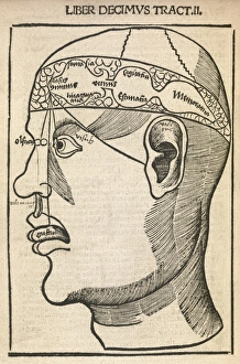
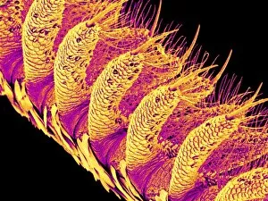
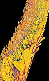
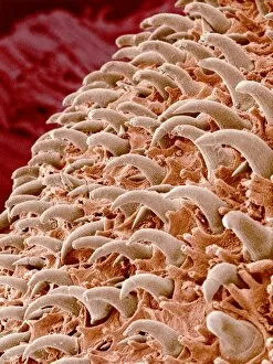
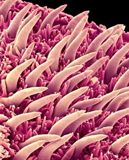
"Exploring the Multifaceted World of Sense: From Motor and Sensory Homunculi to Austen's Characters" In our quest to understand the intricate workings of human perception, we delve into a realm where senses intertwine with art, literature, and science. The concept of "sense" goes beyond mere physicality; it encompasses emotions, intellect, and intuition. From the motor and sensory homunculi that map out our brain's representation of our body parts to Jane Austen's iconic characters in "Sense and Sensibility, " we witness how sense shapes our understanding of ourselves and others. Just as Elinor Dashwood navigates her world guided by reason while Marianne Brandon follows her heart, we too grapple with finding the right balance between logic and emotion. At the University of Oxford's College, scholars have long pondered over Descartes' optics theory from the 17th century. They explore how light interacts with our eyes' intricate anatomy through mesmerizing artwork that captures both its beauty and complexity. Meanwhile, at St Davids College in Lampeter, Cardiganshire, a different kind of engraving adorns its walls – one that depicts Ruthin Castle in Denbighshire. This juxtaposition reminds us that sense is not limited to intellectual pursuits but also embraces nature's grandeur. As we zoom closer into this captivating world of perception, an electron microscope reveals astonishing details about a moth proboscis. Its delicate structure serves as a reminder that even seemingly insignificant creatures possess their own unique ways of sensing their environment. Beyond literature or scientific exploration lies Washington D. C. 's Senate Chamber within Capitol Hill—a place where decisions are made based on political acumen rather than personal sentiments. Here again, sense takes on new dimensions as power dynamics shape collective choices for better or worse. Finally, Eastern Geelong bathes in vibrant colors captured by a photograph—reminding us that they are be a visual feast, evoking emotions and memories with every hue.