
Sex Cell Collection (page 3)
"Exploring the Intricate World of Sex Cells: A Journey through SEM Images" Delve into the mesmerizing realm of sex cells as we embark on a captivating journey


All Professionally Made to Order for Quick Shipping
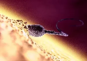
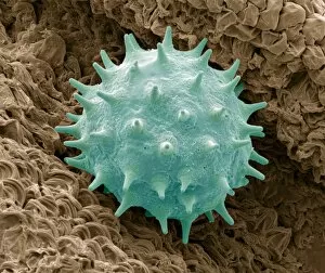
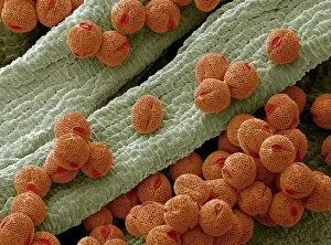
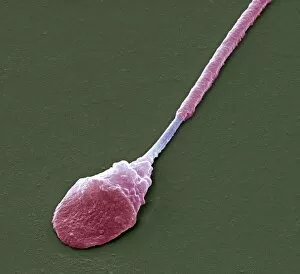
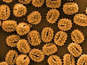
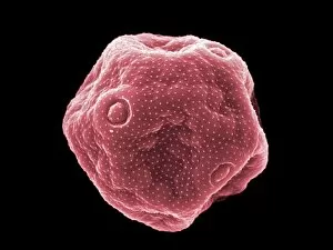
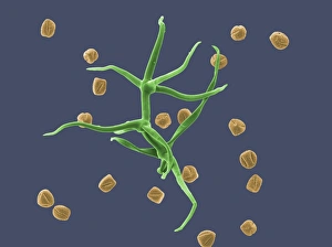
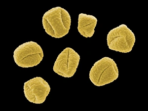
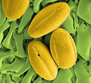
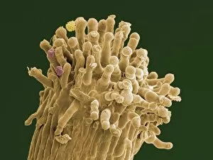
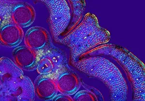
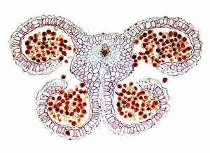
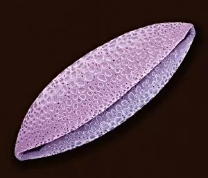
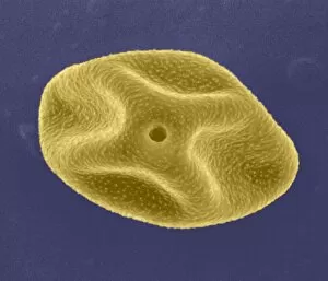
"Exploring the Intricate World of Sex Cells: A Journey through SEM Images" Delve into the mesmerizing realm of sex cells as we embark on a captivating journey, guided by high-resolution scanning electron microscope (SEM) images. Witness the intricate beauty and diversity that lies within these tiny reproductive units. Marvel at the Geranium anther, where delicate pollen grains are nestled in vibrant hues, ready to embark on their crucial mission of fertilization. The Passion flower pollen reveals its unique structure under SEM, showcasing its fascinating intricacies. Observe the Geranium pollen grain with its distinct features, highlighting nature's attention to detail. The Dandelion pollen grain captivates with its spherical shape and textured surface when examined closely under SEM. Discover how pollination occurs as you observe Pollen on a bee leg captured in stunning detail using SEM technology. Philadelphia fleabane pollen grains reveal themselves like miniature jewels upon closer inspection. Witness Gorse stigma adorned with numerous strikingly beautiful pollen grains through SEM imagery. A Lily pollen grain finds itself delicately perched upon a rosemary leaf, creating an enchanting contrast between textures and colors. The Forsythia pollen grains showcase their unique shapes and patterns when magnified using SEM techniques. Chickweed's microscopic yet elegant-looking pollen grains come alive in this visual exploration. Uncover the Hellebore's exquisite morphology through detailed SEM imaging—a testament to nature's artistry even at such minuscule scales. Finally, witness germinating lily pollen as it embarks on new life journeys under close scrutiny from our powerful microscope lenses. Embark on this captivating voyage into the world of sex cells—where beauty meets science—and marvel at nature's ingenuity revealed through astonishingly detailed SEM images capturing every intricate facet of these vital reproductive elements.