
Single Cell Collection
"Exploring the Intricate World of Single Cells: From Acrosphaera Radiolarian to Euastrum Desmid" In the vast realm of microscopic life


All Professionally Made to Order for Quick Shipping
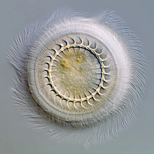
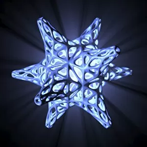
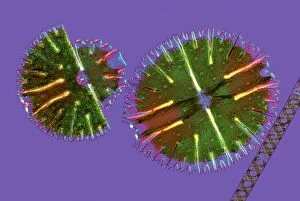
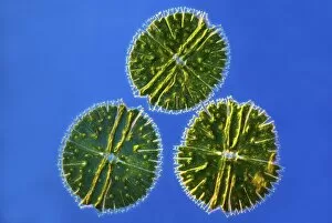
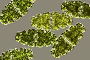
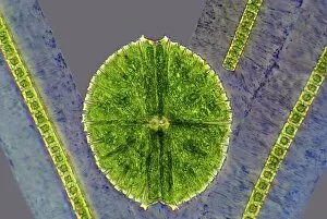
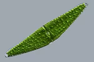
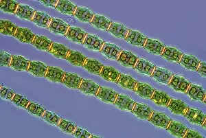
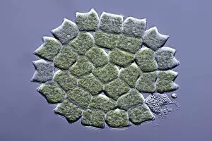
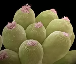
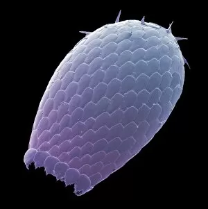
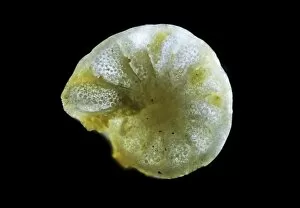
"Exploring the Intricate World of Single Cells: From Acrosphaera Radiolarian to Euastrum Desmid" In the vast realm of microscopic life, single cells hold a mesmerizing beauty that often goes unnoticed. Take a closer look through the lens of science, and you'll discover an enchanting universe teeming with intricate structures and fascinating organisms. One such marvel is Acrosphaera radiolarian, a single-celled creature whose delicate form resembles an ethereal sculpture. Seen under scanning electron microscopy (SEM), its intricately patterned exoskeleton captivates with its symmetrical elegance. On another front, Trichodina parasite reveals itself in a light micrograph. This tiny organism attaches itself to fish gills, showcasing its unique shape and structure while reminding us of nature's complex interconnections. Artistic interpretations also shed light on the captivating world of single-cell lifeforms. The artwork F005 / 0194 depicts the exquisite skeleton of a radiolarian—a testament to nature's ability to create breathtaking designs even at this minuscule scale. Similarly, artwork F005 / 0190 showcases yet another radiant radiolarian skeleton that leaves us in awe. Moving beyond radiolarians, Micrasterias desmids come into focus under light micrographs C016 / 9598 and C016 / 9597. These jewel-like algae exhibit stunning geometric patterns reminiscent of stained glass windows—nature's own masterpieces created by individual cells working harmoniously together. Desmids and spirogyra intertwine in another captivating image captured under light micrograph C016 / 9595. The vibrant green hues blend seamlessly as these single-celled organisms showcase their remarkable adaptability within their aquatic habitats. Netrium desmid takes center stage in light micrograph C016 / 9591—an elegant cell adorned with fine filaments that allow it to glide gracefully through the water.