Fraud Blocker Skip to main content

Single Celled Collection (page 4)

"Unveiling the Microscopic World: Captivating Single-Celled Organisms Under the SEM" Delicate and intricate

Background imageSingle Celled Collection: Foraminiferans, light micrograp

Foraminiferans, light micrograp
Foraminiferans, polarised light micrograph. Foraminiferans are marine single-celled protists that construct and inhabit shells (tests)

Background imageSingle Celled Collection: Frontonia protozoa, light micrograph

Frontonia protozoa, light micrograph
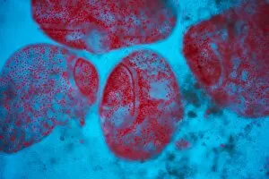
Frontonia protozoa. Light micrograph of a group of Frontonia sp. ciliate protozoa

Background imageSingle Celled Collection: Euglena flagellate protozoan, SEM

Euglena flagellate protozoan, SEM
Euglena flagellate protozoan. Coloured scanning electron micrograph (SEM) of a Euglena sp. flagellate protozoan present in a urine sample taken from someone with a urinary tract infection (UTI)

Background imageSingle Celled Collection: Frontonia protozoan, light micrograph

Frontonia protozoan, light micrograph
Frontonia protozoan. Light micrograph of a Frontonia sp. ciliate protozoan

Background imageSingle Celled Collection: Microsterias green alga, light micrograph

Microsterias green alga, light micrograph
Microsterias green alga. Light micrograph of a Microsterias sp. desmid green algae. Desmids are a common group of unicellular freshwater algae that have intricate cell walls

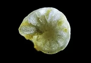
Background imageSingle Celled Collection: Foraminiferan, light micrograph

Foraminiferan, light micrograph. Foraminifera are marine single-celled protozoa that construct and inhabit shells composed of several chambers

Background imageSingle Celled Collection: Pediastrum green algae, light micrograph

Pediastrum green algae, light micrograph
Pediastrum green algae. Light micrograph of a Pediastrum sp. green alga. Members of this genus are non-motile algae that live in freshwater environments

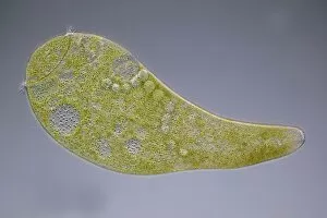
Background imageSingle Celled Collection: Stentor coeruleus protozoan, micrograph

Stentor coeruleus protozoan, micrograph
Stentor coeruleus protozoan. Light micrograph of Stentor coeruleus amongst green algae. This large single-celled protist measures 0.5 to 2 millimetres when fully extended

Background imageSingle Celled Collection: Vorticella protozoan, SEM

Vorticella protozoan, SEM
Vorticella protozoan. Coloured scanning electron micrograph (SEM) of a Vorticella sp. ciliate protozoan. This single-celled organism consists of a bell-shaped head

Background imageSingle Celled Collection: Paramecium protozoan, light micrograph

Paramecium protozoan, light micrograph
Paramecium protozoan. Differential interference contrast micrograph of a Paramecium sp. protozoan. This ciliate protozoan inhabits freshwater, where it feeds mainly on bacteria

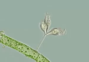
Background imageSingle Celled Collection: Epistylis protozoan colony on algae

Epistylis protozoan colony on algae. Light micrograph of a colony of Epistylis sp. protozoa (centre right) attached to a green algae filament (lower left)

Background imageSingle Celled Collection: Proteus amoeba, light micrograph

Proteus amoeba, light micrograph
Proteus amoeba. Differential interference contrast micrograph of an Amoeba proteus protozoan. This freshwater single-celled organism feeds on bacteria and smaller protozoa

Background imageSingle Celled Collection: Echinocactus pentacanthus cactus, artwork

Echinocactus pentacanthus cactus, artwork
Proteus amoeba. Differential interference contrast micrograph of an Amoeba proteus protozoan. This freshwater single-celled organism feeds on bacteria and smaller protozoa

Background imageSingle Celled Collection: Radiolarian, light micrograph

Radiolarian, light micrograph
Radiolarian, differential interference contrast micrograph. Radiolaria are single-celled protozoans that are found in marine plankton

Background imageSingle Celled Collection: Diatoms, light micrograph

Diatoms, light micrograph. Diatoms are a group of photosynthetic, single-celled algae containing about 10, 000 species. They form an important part of the plankton at the base of the marine

Background imageSingle Celled Collection: Stentor ciliate protozoan, micrograph

Stentor ciliate protozoan, micrograph
Stentor ciliate protozoan. Light micrograph of a Stentor polymorphus ciliate protozoan. This ciliate protozoan inhabits freshwater, where it feeds mainly on bacteria

Background imageSingle Celled Collection: Melosira sp diatoms, light micrograph

Melosira sp diatoms, light micrograph
Diatoms. Light micrograph of Melosira monoliformis freshwater diatoms. Diatoms are a group of photosynthetic, single-celled algae containing about 10, 000 species

Background imageSingle Celled Collection: Pleurosigma sp diatoms, light micrograph

Pleurosigma sp diatoms, light micrograph
Diatoms. Differential interference contrast micrograph of Pleurosigma angulatum marine diatoms. Diatoms are a group of photosynthetic, single-celled algae containing about 10, 000 species

Background imageSingle Celled Collection: Fossil diatoms, light micrograph

Fossil diatoms, light micrograph. Diatoms are a group of photosynthetic, single-celled algae containing about 10, 000 species

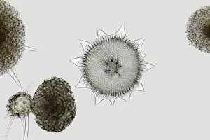
Background imageSingle Celled Collection: Radiolaria, light micrograph

Radiolaria, light micrograph
Radiolaria. Light micrograph of various radiolaria. Radiolaria are single-celled protozoans that are found in marine plankton

Background imageSingle Celled Collection: Stentor ciliate protozoa light micrograph

Stentor ciliate protozoa light micrograph

Background imageSingle Celled Collection: Pleurosigma sp diatom, light micrograph

Pleurosigma sp diatom, light micrograph
Diatom. Light micrograph of Pleurosigma angulatum marine diatoms. Diatoms are a group of photosynthetic, single-celled algae containing about 10, 000 species

Background imageSingle Celled Collection: Macrophage attacking a foreign body, SEM

Macrophage attacking a foreign body, SEM
Macrophage attacking a foreign body. Coloured scanning electron micrograph (SEM) of a macrophage white blood cell (round, left) engulfing a Leishmania mexicana protozoan parasite (long, right)


All Professionally Made to Order for Quick Shipping

"Unveiling the Microscopic World: Captivating Single-Celled Organisms Under the SEM" Delicate and intricate, diatoms reveal their mesmerizing beauty under the scanning electron microscope (SEM). Their symmetrical frustules showcase nature's artistry at its finest. Witness the fascinating process of cell division in yeast cells as they multiply and propagate. The SEM captures this microscopic dance with remarkable detail. Behold the captivating artwork of Trypanosome protozoan, a single-celled organism that evokes both awe and curiosity. Its unique form is brought to life through artistic interpretation. Diatom alga, another marvel of nature, showcases its stunning structure when examined under the SEM lens. These tiny organisms play a crucial role in aquatic ecosystems worldwide. Protozoa, scavengers extraordinaire. Discover these single-celled wonders as they scavenge for particles and microorganisms or absorb nutrients from their environment – truly masters of survival. Acrosphaera radiolarian takes center stage with its intricate skeletal structure captured by SEM imaging techniques. Marvel at this ancient marine creature frozen in time. Journey back millions of years as fossilized diatoms come into focus under the SEM lens, offering glimpses into Earth's past and providing valuable insights for scientific research today. Diatom frustule - an architectural masterpiece on a microscopic scale. Explore these silica-based shells that protect diatoms while showcasing an array of breathtaking patterns through SEM imagery. Oxytricha ciliate protozoan reveals its astonishing complexity when observed using advanced microscopy techniques like SEM C019 / 0253 – unlocking secrets hidden within this enigmatic world. Exploring single-celled organisms through scanning electron microscopy opens up a whole new dimension where beauty meets science; revealing intricacies unseen by our naked eyes but essential to understanding life's diversity on our planet.