
Space Filled Collection (page 3)
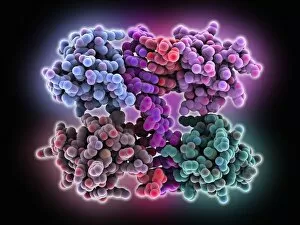
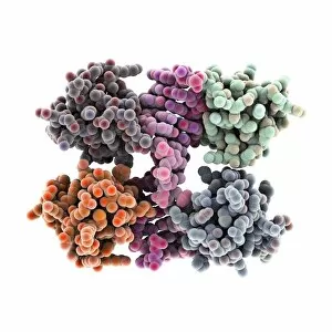
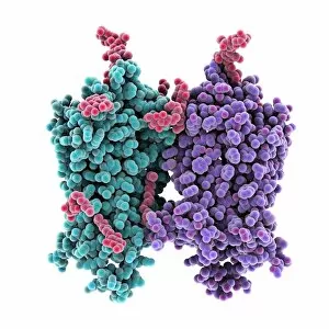
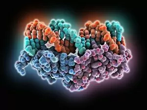
"Space Filled: Exploring the Molecular World" In this captivating image, we delve into the intricate world of molecules


All Professionally Made to Order for Quick Shipping
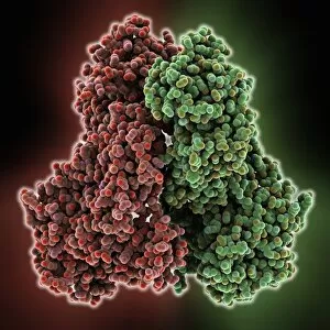
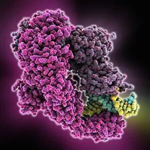
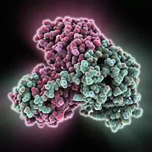
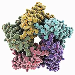
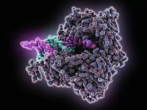
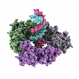
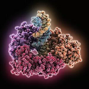
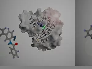
"Space Filled: Exploring the Molecular World" In this captivating image, we delve into the intricate world of molecules, where space is filled with fascinating structures and potential. At the forefront, a remarkable RNA-editing enzyme takes center stage, showcasing its ability to modify genetic information with precision and finesse. Adjacent to it, a molecular model of parathyroid hormone reminds us of its crucial role in regulating calcium levels within our bodies. Its elegant structure hints at the complexity underlying this vital process. Moving further along, we encounter the Donepezil Alzheimer's drug molecule—a powerful compound that aims to combat cognitive decline by inhibiting enzymes responsible for breaking down neurotransmitters. Its compact design reflects its targeted approach towards preserving memory and cognition. Next up is penicillin G—a true game-changer in medicine. This iconic molecule revolutionized antibiotic therapy by targeting bacterial cell walls and effectively treating various infections. Its distinctive shape symbolizes a triumph against microbial adversaries. As we continue our exploration, we come across rosuvastatin—an essential cholesterol-lowering drug that plays a significant role in managing cardiovascular health. The intricacies of its structure hint at how it interacts with specific enzymes involved in cholesterol synthesis. Further on, diclofenac enters the scene as an anti-inflammatory drug renowned for alleviating pain and reducing inflammation caused by conditions such as arthritis or injury. Its dynamic arrangement suggests its ability to target inflammatory pathways precisely. Our journey then takes us to domperidone—an effective anti-sickness medication designed to relieve nausea and vomiting symptoms associated with various conditions. The carefully crafted molecular model showcases how this drug interacts with receptors in our digestive system to restore balance and alleviate discomfort. Lorcaserin emerges next—a promising obesity drug molecule aiming to tackle weight management challenges by suppressing appetite through selective serotonin receptor activation. Its unique architecture alludes to new possibilities for combating obesity-related health concerns. Returning once again is penicillin G—this time, a reminder of its enduring impact on medicine.