Fraud Blocker Skip to main content

Spacefilled Collection (page 5)

"Exploring the Vastness of Spacefilled: From Molecules to Medicines" Delving into the intricate world of molecules, we encounter the Cytochrome b5 molecule C015 / 6696

Background imageSpacefilled Collection: TATA box-binding protein complex C014 / 0879

TATA box-binding protein complex C014 / 0879
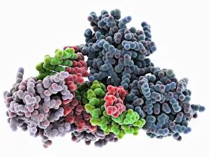
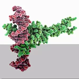
TATA box-binding protein complex. Molecular model showing a TATA box-binding protein (TBP, lilac) complexed with a strand of DNA (deoxyribonucleic acid, green and red)

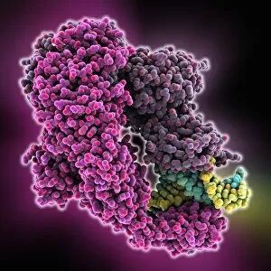
Background imageSpacefilled Collection: Methyladenine glycosylase bound to DNA C014 / 0877

Methyladenine glycosylase bound to DNA C014 / 0877
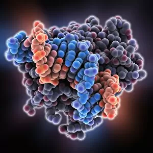
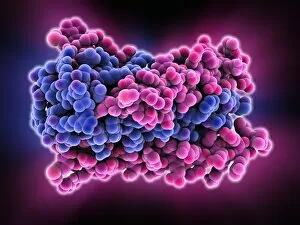
Methyladenine glycosylase bound to DNA. Computer model showing a molecule of human DNA-3-methyladenine glycosylase (purple) in complex with DNA (deoxyribonucleic acid, green and orange)

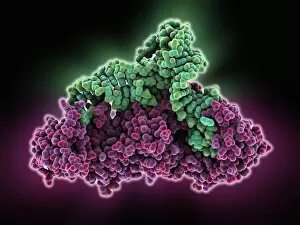
Background imageSpacefilled Collection: Type I topoisomerase bound to DNA C014 / 0883

Type I topoisomerase bound to DNA C014 / 0883
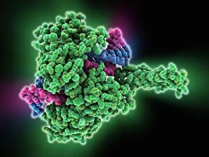
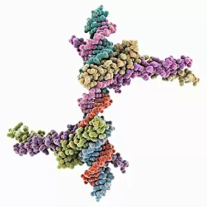
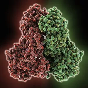
Type I topoisomerase bound to DNA. Molecular model showing a type I topoisomerase molecule (green) bound to a strand of DNA (deoxyribonucleic acid, pink and blue)

Background imageSpacefilled Collection: Resveratrol molecule C014 / 2109

Resveratrol molecule C014 / 2109
Resveratrol, molecular model. Resveratrol is a phytoalexin, a defence chemical produced by plants, found in grapes, peanuts, blueberries and some pines

Background imageSpacefilled Collection: Methyladenine glycosylase bound to DNA

Methyladenine glycosylase bound to DNA. Computer model showing a molecule of human DNA-3-methyladenine glycosylase (purple) in complex with DNA (deoxyribonucleic acid, blue and orange)

Background imageSpacefilled Collection: HIV gp41 glycoprotein C014 / 0866

HIV gp41 glycoprotein C014 / 0866
HIV gp41 glycoprotein. Model showing the molecular structure of the gp41 protein from the HIV (human immunodeficiency virus) glycoprotein envelope

Background imageSpacefilled Collection: Transcription factor complexed with DNA C014 / 0869

Transcription factor complexed with DNA C014 / 0869
Transcription factor complexed with DNA. Computer model showing sterol regulatory element binding transcription factor 1 (SREBF1, horizontal) bound to a section of DNA (deoxyribonucleic acid)

Background imageSpacefilled Collection: Zinc finger bound to DNA C014 / 0864

Zinc finger bound to DNA C014 / 0864
Zinc finger bound to DNA. Molecular model showing a zinc finger molecule bound to a strand of DNA (deoxyribonucleic acid)

Background imageSpacefilled Collection: Epstein-Barr virus protein bound to DNA C014 / 0875

Epstein-Barr virus protein bound to DNA C014 / 0875
Epstein-Barr virus protein bound to DNA. Computer model showing a molecule of Epstein-Barr nuclear antigen 1 (EBNA1) bound to a strand of DNA (deoxyribonucleic acid)

Background imageSpacefilled Collection: HIV gp41 glycoprotein

HIV gp41 glycoprotein. Model showing the molecular structure of the gp41 protein from the HIV (human immunodeficiency virus) glycoprotein envelope

Background imageSpacefilled Collection: ATP synthase molecule

ATP synthase molecule. Molecular model showing the structure of ATP synthase (ATPase) subunit C. ATPase is an important enzyme that provides energy for cells through the synthesis of adenosine

Background imageSpacefilled Collection: Transcription factor complexed with DNA C014 / 0870

Transcription factor complexed with DNA C014 / 0870
Transcription factor complexed with DNA. Computer model showing a max protein (green) bound to a strand of DNA (deoxyribonucleic acid, pink)

Background imageSpacefilled Collection: Nucleosome core particle bound to DNA C014 / 0872

Nucleosome core particle bound to DNA C014 / 0872
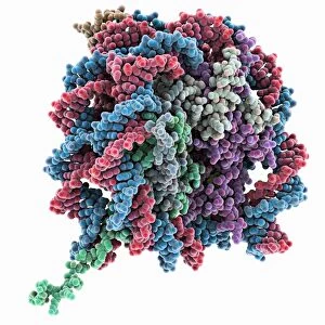
Nucleosome core particle bound to DNA. Molecular model showing a nucleosome core particle (green and purple) bound to a strand of DNA (deoxyribonucleic acid, blue and red)

Background imageSpacefilled Collection: Chaperonin protein complex C014 / 0873

Chaperonin protein complex C014 / 0873
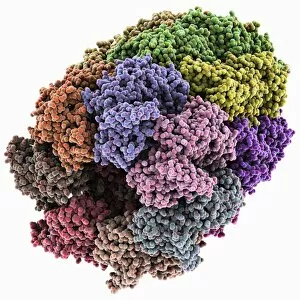
Chaperonin protein complex. Molecular model showing the structure of a GroEL/GroES/(ADP)7 chaperonin complex. Chaperonins are proteins that provide favourable conditions for the correct folding of

Background imageSpacefilled Collection: Chaperonin protein complex

Chaperonin protein complex. Molecular model showing the structure of a GroEL/GroES/(ADP)7 chaperonin complex. Chaperonins are proteins that provide favourable conditions for the correct folding of

Background imageSpacefilled Collection: Coenzyme Q10 molecule C014 / 2294

Coenzyme Q10 molecule C014 / 2294
Coenzyme Q10, molecular model. This is the fully oxidised version of the molecule, known as ubiquinone. Coenzyme Q10 is involved in the electron transport chain that synthesises (ATP)

Background imageSpacefilled Collection: Gabapentin enacarbil drug molecule

Gabapentin enacarbil drug molecule. Computer model showing the structure of a molecule of the anticonvulsant and analgesic (painkilling) drug gabapentin enacarbil (Horizant)

Background imageSpacefilled Collection: Cholecystokinin-8 molecule C014 / 4895

Cholecystokinin-8 molecule C014 / 4895
Cholecystokinin-8 molecule. Computer model showing the structure of the terminal fragment of a molecule of the hormone cholecystokinin-8 (CCK-8)

Background imageSpacefilled Collection: Activated ghrelin hormone molecule C014 / 4903

Activated ghrelin hormone molecule C014 / 4903
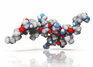
Activated ghrelin hormone molecule. Computer model showing the structure of the human hormone ghrelin. Atoms are colour-coded spheres (carbon: grey, oxygen: red, nitrogen: blue)

Background imageSpacefilled Collection: Obestatin molecule C014 / 4908

Obestatin molecule C014 / 4908
Obestatin molecule. Computer artwork showing the structure of a molecule of obestatin. Obestatin is thought to supress hunger and reduce food intake, thereby reducing weight gain

Background imageSpacefilled Collection: Anthrax lethal factor molecule

Anthrax lethal factor molecule
Anthrax lethal factor, molecular model. This enzyme is one of three protein components that form the anthrax toxin produced by the bacterium Bacillus anthracis

Background imageSpacefilled Collection: E. coli DNA mismatch repair complex

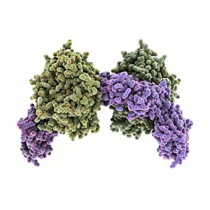
E. coli DNA mismatch repair complex. Computer model showing the structure of a MutS (Mutator S) protein from Escherichia coli (E)

Background imageSpacefilled Collection: Glutaminyl-tRNA synthetase molecule

Glutaminyl-tRNA synthetase molecule
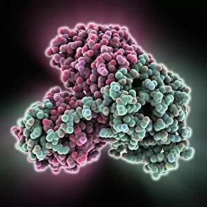
Glutaminyl-tRNA synthetase protein molecule. Molecular model showing bacterial glutaminyl-tRNA synthetase complexed with glutamine tRNA (transfer ribonucleic acid)

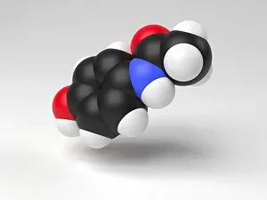
Background imageSpacefilled Collection: Paracetamol molecule

Paracetamol molecule
Serotonin molecule. Computer model showing the structure of a molecule of the neurotransmitter (nerve signalling chemical) serotonin (5-hydroxytryptamine)

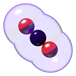
Background imageSpacefilled Collection: Carbon dioxide molecules C016 / 8495

Carbon dioxide molecules C016 / 8495
Carbon dioxide molecules. Computer artwork showing the structure of a molecule of carbon dioxide. Carbon dioxide is a colourless gas that occurs naturally in the atmosphere

Background imageSpacefilled Collection: Carbon dioxide molecule C016 / 8494

Carbon dioxide molecule C016 / 8494
Carbon dioxide molecule. Computer artwork showing the structure of a molecule of carbon dioxide. Carbon dioxide is a colourless gas that occurs naturally in the atmosphere

Background imageSpacefilled Collection: Carbon dioxide molecule C016 / 8493

Carbon dioxide molecule C016 / 8493
Carbon dioxide molecule. Computer artwork showing the structure of a molecule of carbon dioxide. Carbon dioxide is a colourless gas that occurs naturally in the atmosphere

Background imageSpacefilled Collection: Bacterial RNA-binding protein molecule

Bacterial RNA-binding protein molecule
Bacterial RNA-binding protein. Molecular model of the bacterial RNA-binding protein Hfq complexed with RNA (ribonucleic acid)

Background imageSpacefilled Collection: Nucleoplasmin molecule

Nucleoplasmin molecule
Nucleoplamsin, molecular model. This protein is involved in nucleosome assembly. Nucleosomes are the fundamental repeating unit used to package DNA (deoxyribonucleic acid) inside cell nuclei

Background imageSpacefilled Collection: Vitamin B1 molecule C016 / 8278

Vitamin B1 molecule C016 / 8278
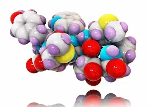
Vitamin B1 molecule. Computer model showing the structure of a molecule of vitamin B1 (thiamine). Atoms are represented as colour-coded spheres: carbon (light blue), hydrogen (white)

Background imageSpacefilled Collection: Vitamin B1 molecule C016 / 8276

Vitamin B1 molecule C016 / 8276
Vitamin B1 molecule. Computer model showing the structure of a molecule of vitamin B1 (thiamine). Vitamin B1 is an essential nutrient that humans are unable to produce

Background imageSpacefilled Collection: cAMP-dependent protein kinase molecule C016 / 2063

cAMP-dependent protein kinase molecule C016 / 2063
cAMP-dependent protein kinase, molecular model. This enzyme is also known as protein kinase A (PKA). This is the holoenzyme, which consists of two regulatory subunits and two catalytic subunits

Background imageSpacefilled Collection: Bacterial transcription factor molecule C016 / 2061

Bacterial transcription factor molecule C016 / 2061
Bacterial transcription factor. Molecular model of the transcription factor AmrZ from the bacterium Pseudomonas aeruginosa complexed with DNA (deoxyribonucleic acid)

Background imageSpacefilled Collection: Packaged DNA molecule

Packaged DNA molecule. Computer model showing the structure of a smoothly bent molecule of DNA (deoxyribonucleic acid) as it would appear in chromatin

Background imageSpacefilled Collection: MHC protein-antigen complex

MHC protein-antigen complex. Computer model showing a histocompatibility antigen complexed to a class II MHC (major histocompatibility complex) protein molecule

Background imageSpacefilled Collection: DNA mismatch repair complex

DNA mismatch repair complex. Computer model showing the structure of a MutS (Mutator S) protein binding to DNA (deoxyribonucleic acid) during DNA mismatch repair

Background imageSpacefilled Collection: Human electron transfer flavoprotein

Human electron transfer flavoprotein. Computer model showing the structure of a human electron transfer flavoprotein (ETF) molecule

Background imageSpacefilled Collection: Ribosomal RNA-binding protein molecule

Ribosomal RNA-binding protein molecule. Computer model showing the structure of a ribosomal protein L9 (RPL9) molecule from Bacillus stearothermophilus bacteria

Background imageSpacefilled Collection: Signal recognition particle RNA molecule

Signal recognition particle RNA molecule. Computer model showing the molecular structure of the 2 A structure of helix 6 of the human signal recognition particle (SRP) RNA (ribonucleic acid)

Background imageSpacefilled Collection: Synthetic DNA triplex molecule

Synthetic DNA triplex molecule. Computer model showing the molecular structure of a synthetic intramolecular DNA (deoxyribonucleic acid) triplex linked by hexakis(ethylene glycol) units

Background imageSpacefilled Collection: Ebola virus glycoprotein

Ebola virus glycoprotein. Computer model showing the structure of a GP2 glycoprotein from the outer envelope of an Ebola virus (EBOV)

Background imageSpacefilled Collection: HP1 molecule C-terminal domain

HP1 molecule C-terminal domain. Computer model showing the structure of the C terminal (shadow chromo) domain of a heterochromatin protein 1 (HP1) molecule from a mouse


All Professionally Made to Order for Quick Shipping

"Exploring the Vastness of Spacefilled: From Molecules to Medicines" Delving into the intricate world of molecules, we encounter the Cytochrome b5 molecule C015 / 6696, a key player in electron transfer processes within cells. Unraveling the mysteries of DNA structure, we come across the Z-DNA tetramer molecule C015 / 6557, showcasing its unique left-handed helical conformation. Embarking on a psychedelic journey, we discover the Psilocybin drug molecule, known for its mind-altering effects and potential therapeutic applications. Witnessing precision at work, we examine the RNA-editing enzyme molecular model that plays a crucial role in modifying genetic information to ensure cellular functionality. Energizing our bodies from within, we explore the ATPase molecule responsible for powering various cellular processes by converting ATP into ADP and phosphate. Battling against viral invaders, we observe Rhinovirus and antibody molecular models (C015 / 7139 & C015 / 7138) engaged in an intricate dance of recognition and defense. Admiring art imitating life, we marvel at an artwork depicting DNA's elegant double helix structure (C017 / 7217), symbolizing life's blueprint encoded within our genes. Nourishing our bodies with essential building blocks, Valine molecule takes center stage as one of the amino acids vital for protein synthesis and muscle repair. Embracing metabolic pathways with Methionine molecule as our guide; this sulfur-containing amino acid is indispensable for protein synthesis and methylation reactions. Discovering Histidine's multifaceted nature - not only serving as an amino acid but also playing pivotal roles in pH regulation and metal ion coordination within proteins.