Fraud Blocker Skip to main content

Spongy Collection

"Spongy: Exploring the Intricate World of Bone Structure and Beyond" In this captivating journey

Background imageSpongy Collection: Osteoporotic bone, SEM

Osteoporotic bone, SEM
Osteoporotic bone. Coloured scanning electron micrograph (SEM) of fractured bone tissue from a patient suffering from osteoporosis (brittle bone disease)

Background imageSpongy Collection: Picture No. 11675585

Picture No. 11675585
Scanning Electron micrograph (SEM)showing stomata on a Yew Leaf. Date:

Background imageSpongy Collection: Osteoporotic bone

Osteoporotic bone

Background imageSpongy Collection: Sycon and Aplysina, No. 12, Pfurtschellers Zoological Wall Chart (colour litho)

Sycon and Aplysina, No. 12, Pfurtschellers Zoological Wall Chart (colour litho)
HMB306153 Sycon and Aplysina, No.12, Pfurtschellers Zoological Wall Chart (colour litho) by Pfurtscheller, Paul (1855-1927); 130x122 cm; Humboldt-Universitaet, Berlin

Background imageSpongy Collection: Root nodule

Root nodule. Coloured scanning electron micrograph (SEM) of a root nodule on a pea plant (Pisum sativum) caused by the nitrogen-fixing soil bacteria Rhizobium leguminosarum

Background imageSpongy Collection: Cross-section diagram of a human long bone

Cross-section diagram of a human long bone

Background imageSpongy Collection: Picture No. 11675968

Picture No. 11675968
Spongy Green Moss over lava fields. Date:

Background imageSpongy Collection: Cross-section of spongy fruit pie, close up

Cross-section of spongy fruit pie, close up

Background imageSpongy Collection: A natural sponge

A natural sponge

Background imageSpongy Collection: Human bone structure, artwork C016 / 7494

Human bone structure, artwork C016 / 7494
Human bone structure. Computer artwork showing all the different parts that make up a human bone, including the bone marrow (bottom left) where blood cells (round, white and red) are formed

Background imageSpongy Collection: Cancellous bone, light micrograph C016 / 0509

Cancellous bone, light micrograph C016 / 0509
Cancellous bone. Light micrograph of a section through cancellous, or spongy, bone. Constructed in a honeycomb-type architecture

Background imageSpongy Collection: Chimpanzee skull, X-ray

Chimpanzee skull, X-ray
Chimpanzee skull. Coloured X-ray of a side view of a chimpanzees (Pan troglodytes) skull. The cranium (upper right) is considerably smaller than in humans

Background imageSpongy Collection: Flys proboscis, SEM

Flys proboscis, SEM
Flys proboscis. Coloured scanning electron micrograph (SEM) of the tip of the proboscis (snout) of an unidentified fly (order Diptera). Flies possess mouthparts adapted for piercing and sucking

Background imageSpongy Collection: Flys proboscis, light micrograph

Flys proboscis, light micrograph
Flys proboscis. Light micrograph (LM) of the tip of the proboscis (snout) of a fly (order Diptera). Flies possess mouthparts adapted for piercing and sucking

Background imageSpongy Collection: Fly proboscis, SEM

Fly proboscis, SEM
Fly proboscis. Coloured scanning electron micrograph (SEM) of the tip of the proboscis (snout) of an unidentified fly (order Diptera). Flies possess mouthparts adapted for piercing and sucking

Background imageSpongy Collection: Osteoclast, TEM

Osteoclast, TEM
Osteoclast. Coloured transmission electron micrograph (TEM) of an osteoclast (blue and purple). Osteoclasts are found in cavities, known as Howslips lacunae, on the surface of bones

Background imageSpongy Collection: Bone, SEM

Bone, SEM
Spongy bone. Coloured scanning electron micrograph (SEM) of freeze-fractured spongy bone tissue. It shows the matrix of collagen and calcium crystals that form the structure of bone tissue

Background imageSpongy Collection: Bone anatomy

Bone anatomy. Computer artwork showing the anatomy of a normal bone shaft. An outer layer of compact bone is made up of cylindrical units (osteons, black stripes)

Background imageSpongy Collection: Spongy bone, SEM

Spongy bone, SEM
Spongy bone. Coloured scanning electron micrograph (SEM) of human spongy (cancellous) bone from the shaft of a long bone. Found inside bones, below a dense shell of hard compact bone

Background imageSpongy Collection: Bone growth, light micrograph

Bone growth, light micrograph
Bone growth. Light micrograph of actively growing cells in the epiphyseal plate (growth plate) between the diaphysis (shaft) and epiphysis (rounded end) of a long bone

Background imageSpongy Collection: Interior of a bone, SEM

Interior of a bone, SEM
Interior of a bone. Coloured scanning electron micrograph (SEM) of a long bone fractured to show its interior. The outer layer (pink) of the bone comprises proliferating cartilage

Background imageSpongy Collection: Household sponge, SEM

Household sponge, SEM
Household sponge. Coloured scanning electron micrograph (SEM) of a household sponge. Its porous structure enables it to retain liquid. Magnification unknown

Background imageSpongy Collection: Bird bone, SEM

Bird bone, SEM
Bird bone. Coloured scanning electron micrograph (SEM) of the interior of bone from a birds skull. This porous structure found inside bone is called cancellous (spongy) bone

Background imageSpongy Collection: Osteoporotic bone, artwork

Osteoporotic bone, artwork
Osteoporosis. Computer artwork of osteoporotic bone. Osteoporosis (brittle bone disease) causes a reduction in overall bone mass and an increase in the bones porosity

Background imageSpongy Collection: Soft rush stem, light micrograph

Soft rush stem, light micrograph
Soft rush stem. Fluorescent light micrograph of a cross section through pith from a soft rush (Juncus effusus) stem. The star-shaped aerenchyma tissue (upper frame)

Background imageSpongy Collection: Bogbean stem, light micrograph

Bogbean stem, light micrograph
Bogbean stem. Light micrograph of a cross-section through the stem of a bogbean plant (Menyanthes trifoliata), showing its internal structure


All Professionally Made to Order for Quick Shipping

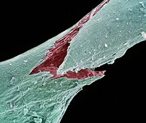
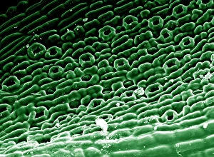
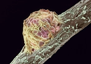
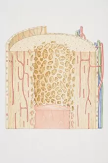
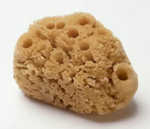
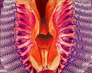
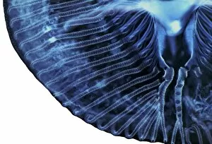
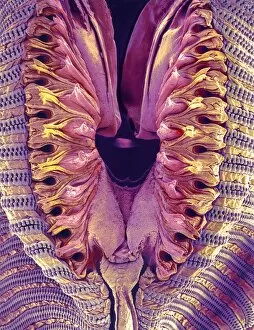
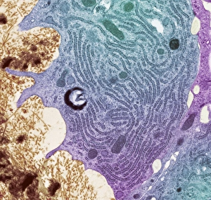
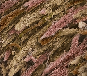
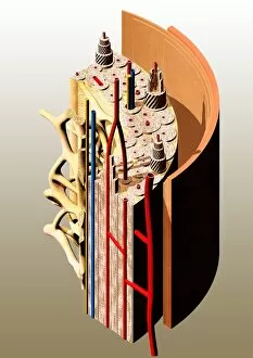
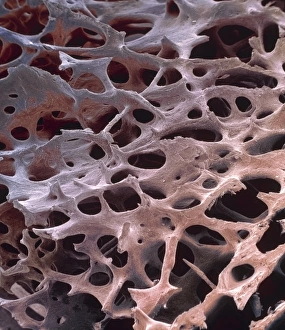
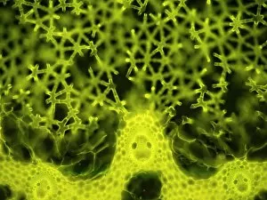
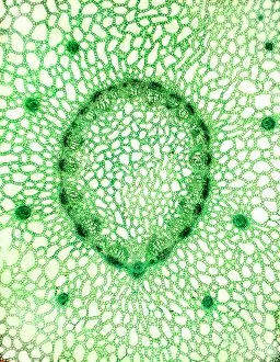
"Spongy: Exploring the Intricate World of Bone Structure and Beyond" In this captivating journey, we delve into the fascinating realm of "spongy" - a term that takes us from osteoporotic bone to natural sponges, unveiling hidden wonders along the way. Starting with an image captured by a scanning electron microscope (SEM), Picture No. 11675585 reveals the intricate details of osteoporotic bone, showcasing its porous nature. This visual representation serves as a reminder of the fragility and importance of maintaining healthy bones. Moving on to Pfurtschellers Zoological Wall Chart, we encounter Sycon and Aplysina in Picture No. 12 - vibrant organisms that resemble sponges but exist in aquatic ecosystems. Their unique structures remind us that sponginess can be found not only within our bodies but also in diverse forms throughout nature. A cross-section diagram of a human long bone depicted in Picture No. 11675968 provides further insight into the complex architecture beneath our skin's surface. The interplay between compact and cancellous bone highlights how both strength and flexibility are crucial for optimal skeletal function. Shifting gears towards culinary delights, we encounter a mouthwatering close-up shot - Cross-section fruit pie. This delectable treat showcases how even food can possess elements reminiscent of sponge-like textures, inviting us to explore unexpected connections between taste sensations and anatomical features. Venturing back into nature's realm, we come across an awe-inspiring sight - a natural sponge floating effortlessly underwater. Its delicate yet resilient structure reminds us that beauty often lies within simplicity while offering inspiration for innovative materials design beyond traditional applications. Artwork C016 / 7494 presents Human Bone Structure as an artistic interpretation capturing the essence of our inner framework with striking visuals that merge science and creativity seamlessly together. Delving deeper into microscopic realms through light micrograph C016 / 0509, we encounter cancellous bone.