Fraud Blocker Skip to main content

Stain Collection (page 16)

"Unveiling the Intricacies: A Stain's Tale" In the vast realm of microscopic wonders, a stain reveals hidden secrets within cerebellum tissue

Background imageStain Collection: Actin tail, fluorescent micrograph

Actin tail, fluorescent micrograph
Actin tail. Immunofluorescence deconvolution micrograph of a close-up of an actin filament (green) in a cell infected with vaccinia virus particles

Background imageStain Collection: Vaccinia virus infected cell

Vaccinia virus infected cell. Immunofluorescence deconvolution micrograph of a cell infected with vaccinia virus particles. Host and viral DNA (deoxyribonucleic acid) is blue

Background imageStain Collection: E. coli infection mechanism

E. coli infection mechanism. Immunofluorescence deconvolution micrograph of Escherichia coli bacteria (centre). Bacterial DNA (deoxyribonucleic acid) is blue

Background imageStain Collection: Fibroblast cells, fluorescent micrograph

Fibroblast cells, fluorescent micrograph
Fibroblast cells. Immunofluorescence deconvolution micrograph of fibroblast cells. The cell nuclei, which contain the cells genetic information, are blue

Background imageStain Collection: Maize niblet, light micrograph

Maize niblet, light micrograph
Maize niblet. Light micrograph of a maize niblet, the seed of the maize plant (Zea mays). A seed contains an embryo, a fertilised ovule from which a new plant can grow (green, upper left)

Background imageStain Collection: Wheat stem, light micrograph

Wheat stem, light micrograph
Wheat stem. Light micrograph of a section through the stem of a wheat grass (Triticum aestivium). The circular structures (orange and green) are vascular bundles

Background imageStain Collection: Oviduct mucosal folds, light micrograph

Oviduct mucosal folds, light micrograph. This section through the folds shows the ciliated columnar epithelium (red) and the connective tissue core (green)

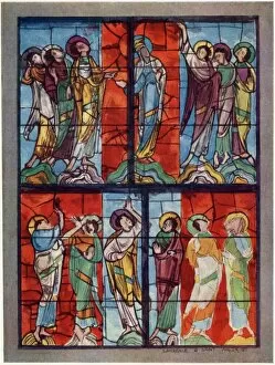
Background imageStain Collection: Stain Glass at Le Mans

Stain Glass at Le Mans
The Apostles watch the ascension of Jesus in this window at Le Mans, France, supposed to date from the 13th century

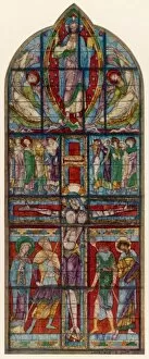
Background imageStain Collection: Stain Glass Crucifixion

Stain Glass Crucifixion
A magnificent representation of Jesuss crucifixion in a brightly coloured window at Poitiers, France

Background imageStain Collection: Stain Glass Rouen

Stain Glass Rouen
The life of Saint Gervais is depicted in a series of panels in this window at Saint Ouen, Rouen, France

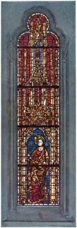
Background imageStain Collection: Stain Glass York Minster

Stain Glass York Minster
The figure of Saint Margaret depicted in the west window of the north aisle of York minster

Background imageStain Collection: Cup of coffee

Cup of coffee placed on top of a bill

Background imageStain Collection: Stain Glass Chartres

Stain Glass Chartres
This window at Chartres, France, depicts Amaury de Montfort, a celebrated French noble, though it could be anyone, really


All Professionally Made to Order for Quick Shipping

"Unveiling the Intricacies: A Stain's Tale" In the vast realm of microscopic wonders, a stain reveals hidden secrets within cerebellum tissue, captured in a mesmerizing light micrograph. As we delve into this captivating world, echoes of Sherlock Holmes' thrilling adventure "The Adventure of the Second Stain" resonate. On the title-page of Thomas Paine's pamphlet Common Sense, owned by John Adams, a faint yet significant mark hints at an untold story. Just like pine pollen grains and pine stem depicted in delicate light micrographs, this stain holds clues waiting to be deciphered. As our journey continues through intricate lime tree stems and cerebral cortex nerve cells, we find ourselves transported to Baker Street alongside Holmes and Watson. The enigmatic duo unravels mysteries that lie beneath seemingly ordinary surfaces – just as stains reveal hidden truths. Within the maize root's vibrant structure lies another glimpse into the microscopic world. Light micrographs capture its essence with utmost precision while reminding us that even nature conceals its own stories within each stain. Returning full circle to cerebellum tissue once more, we realize how these stains connect diverse realms – from literature to science – weaving together tales both fictional and factual. Each stain represents a unique narrative waiting for curious minds to explore their depths. Just like Holmes' keen eye for detail or an astute scientist peering through a microscope lens, let us embrace these stains as gateways into uncharted territories. In every speckle or smudge lies an opportunity for discovery - an invitation to uncover secrets that may reshape our understanding of the world around us. So let us embark on this extraordinary odyssey where stains become storytellers themselves; where adventures await those who dare to look beyond what meets the eye; where even something as simple as a blot can hold profound significance in unraveling life's grand tapestry.