
Striae Collection
"Exploring the Intricate World of Striae: Unveiling Diatom Structures through SEM Imaging" Diatoms, a type of microscopic algae


All Professionally Made to Order for Quick Shipping
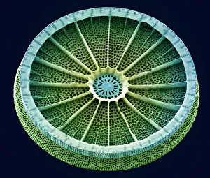
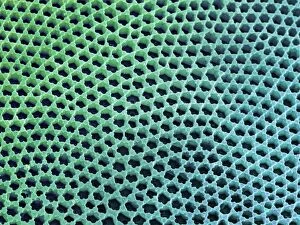
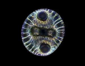
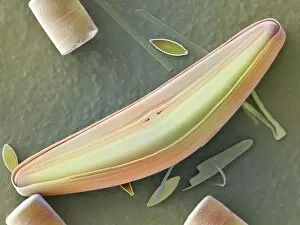
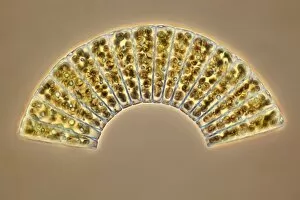
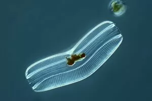
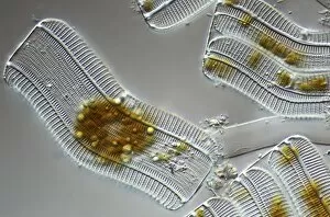
"Exploring the Intricate World of Striae: Unveiling Diatom Structures through SEM Imaging" Diatoms, a type of microscopic algae, have long fascinated scientists with their intricate cell walls known as frustules. Using Scanning Electron Microscopy (SEM), researchers can delve into the mesmerizing details of these diatom shells and uncover the beauty hidden within. In this captivating image captured by SEM, we witness the remarkable structure of a diatom cell wall. The delicate striae, or fine lines, etched on its surface create an exquisite pattern that resembles nature's own artwork. These striae serve various functions for diatoms – from providing strength to facilitating nutrient absorption. Zooming in further with SEM reveals even more astonishing features. Tabellaria diatoms take center stage in another striking image (C016 / 9599). Their elongated shape and meticulously arranged striae showcase the diversity within this fascinating group of organisms. But it's not just living diatoms that captivate our attention; fossilized remains also offer glimpses into ancient ecosystems. A stunning light micrograph (C016 / 8603) showcases a fossilized diatom, frozen in time yet preserving its intricate structures for us to marvel at. Returning to SEM imaging, we encounter Tabellaria diatoms once again (C016 / 9600). This time, their unique arrangement creates an almost symmetrical pattern reminiscent of kaleidoscopic artistry. Each tiny detail contributes to the overall aesthetic appeal and scientific significance of these organisms. As we continue our journey through the world using advanced microscopy techniques like SEM and light micrographs such as C014 / 4673, we gain a deeper appreciation for the complexity and elegance found within diatoms' microscopic realm. These images remind us that even at scales invisible to the naked eye, nature never fails to astound us with its intricacy and beauty.