
Support Cell Collection
"Unveiling the Intricate World of Support Cells: A Journey Through Micrographs and Artwork" Glial cells


All Professionally Made to Order for Quick Shipping
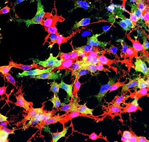
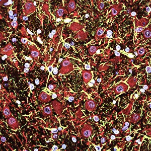
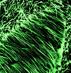
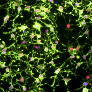
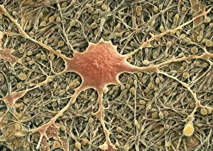
"Unveiling the Intricate World of Support Cells: A Journey Through Micrographs and Artwork" Glial cells, confocal light micrograph: Delving into the hidden network of support cells in our brain, glial cells take center stage in this captivating confocal light micrograph. Glial stem cell culture, light micrograph: Witness the birthplace of future support cells as you explore this mesmerizing light micrograph showcasing a glial stem cell culture. Cerebral cortex nerve cells: Behold the intricate interplay between nerve cells and their faithful companions - support cells - within the cerebral cortex, captured in stunning detail. Cerebellum tissue, light micrograph: Step into the world of balance and coordination as you marvel at this breathtaking light micrograph unveiling the complex relationship between cerebellum tissue and its supporting cast of glial cells. Myelin sheaths and glial cells, artwork C014 / 2646 & 2647: Immerse yourself in an artistic representation that beautifully illustrates how myelin sheaths intertwine with diligent glial cells to safeguard our nerves' integrity. Retina, light micrograph: Explore the delicate intricacies of vision through this illuminating light micrograph capturing support cell activity within our retina – a testament to their crucial role in maintaining visual function. Nerve support cell, SEM (Scanning Electron Microscope): Peer through a microscopic lens to witness a nerve support cell like never before – its intricate structure magnified by cutting-edge technology for your amazement. Muscle motor neurons, light micrograph: Discover how muscle motor neurons rely on steadfast support from specialized cellular allies showcased vividly in this enlightening light micrograph study. Motor neurons, light micrograph: Embark on an exploration deep within our nervous system as you encounter motor neurons harmoniously interacting with their support cells, captured in striking detail.