
Synapse Collection
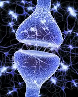
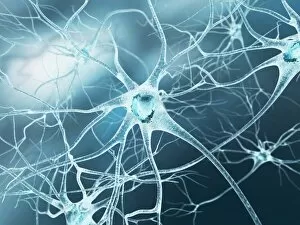
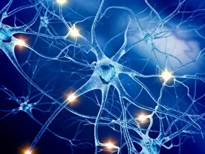
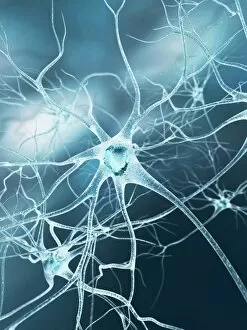
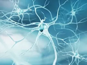
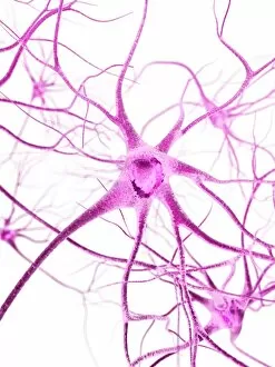
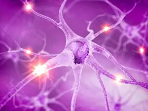
A synapse, also known as a nerve junction, is a crucial element in the intricate network of our nervous system


All Professionally Made to Order for Quick Shipping
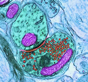
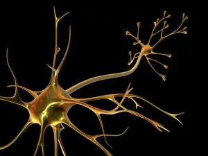
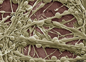
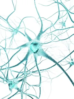
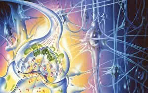
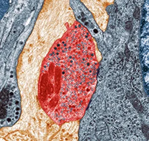
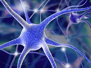
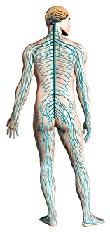
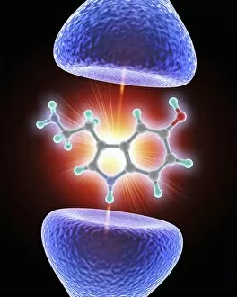
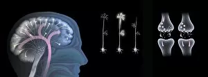
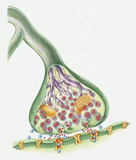
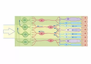
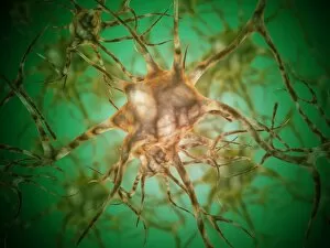
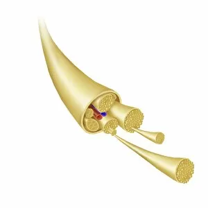
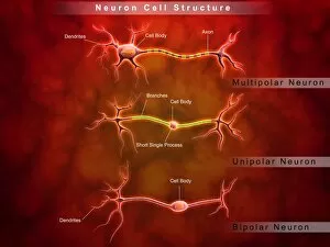
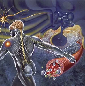
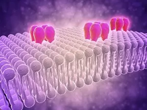
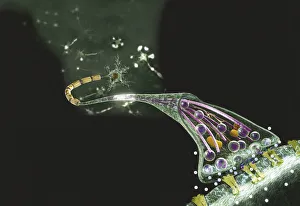
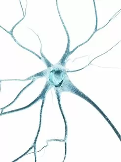
A synapse, also known as a nerve junction, is a crucial element in the intricate network of our nervous system, and is through these synapses that information travels between nerve cells, allowing us to perceive and react to the world around us. Using advanced imaging techniques like Transmission Electron Microscopy (TEM) and Scanning Electron Microscopy (SEM), scientists have been able to capture detailed images of these synapses. In one such image, an artistic representation of a nerve cell called artwork F007/7448 showcases the complexity and beauty of this microscopic structure. Another TEM image reveals the intricacy of a nerve synapse at an even closer level. The delicate connections between neurons can be seen in stunning detail, highlighting their importance in transmitting signals throughout our body. Computer-generated artwork depicting multiple nerve cells further emphasizes the vastness and interconnectedness of our neural network. These illustrations serve as visual reminders that every thought, movement, or sensation we experience relies on countless synapses working together seamlessly. In addition to capturing static images, diagrams provide valuable insights into how our nervous system functions. A posterior view diagram illustrates the human nervous system's layout while shedding light on its role in coordinating bodily functions. The effects of ecstasy on brain function are another area where understanding synaptic activity becomes crucial. Research has shown that this recreational drug alters neurotransmitter levels within synapses, leading to various physiological and psychological changes. Cross-section illustrations offer yet another perspective on synapses' inner workings. Biomedical depictions reveal neurotransmitters crossing from neuron to target cell—a vital process for relaying messages across different parts of our body effectively. Ultimately, exploring these cross-sections provides invaluable knowledge about how nerves communicate with each other via synaptic connections—knowledge that paves the way for advancements in neuroscience research and potential treatments for neurological disorders.