
Tissues Collection
Tissues: Unveiling the Hidden World within Our Bodies Step into a world of wonder as we explore the intricate and diverse realm of tissues


All Professionally Made to Order for Quick Shipping
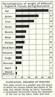
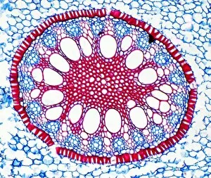
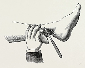
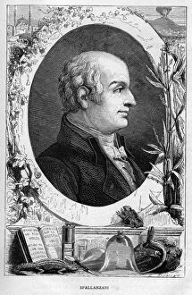
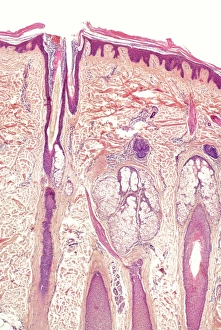
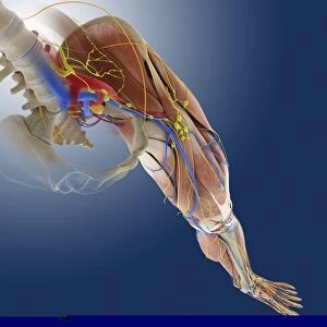
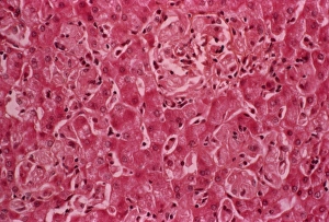
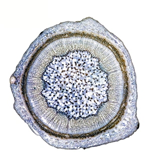
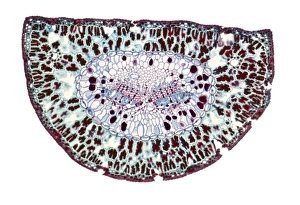
Tissues: Unveiling the Hidden World within Our Bodies Step into a world of wonder as we explore the intricate and diverse realm of tissues. Just like a teddy bear concealing hidden objects, our bodies are filled with these remarkable structures that play vital roles in our health and well-being. Under the microscope, we discover mesmerizing images that showcase nature's artistry. The maize root, elm stem, and dog rose stem light micrographs reveal an astonishing complexity within plant tissues. Each delicate cell intertwines to support growth and nourishment, reminding us of the interconnectedness of all living things. But it is not just plants that possess fascinating tissue compositions; humans too have their own unique tapestry. Starvation leaves its mark on our bodies, degrees of wasting captured lithographically showcasing the heartbreaking consequences when they can deprived of sustenance. In contrast to this somber reality, a black-and-white photo captures a figure engaging in mechanical exercise—a testament to human resilience and determination to keep our tissues strong through physical activity. Art also finds inspiration in tissues. Hokusai's "Horse Pattern" from his renowned series "Horses" showcases how even ancient artists recognized the beauty found within these biological wonders. Beyond aesthetics lies innovation—tissue manufacture revolutionizes medical advancements by providing life-saving solutions for those in need. Iris root light micrograph reminds us that progress often stems from understanding nature at its core. Yet sometimes circumstances demand more drastic measures—the mode of dividing tissues during amputation requires specialized medical equipment and surgical instruments designed with precision to ensure patient well-being while preserving functionality. As we delve deeper into this captivating subject matter, Picture No. 10877009 transports us back in time—to Lazzaro Spallanzani's groundbreaking work disproving spontaneous generation theory—an essential milestone in unraveling the mysteries surrounding bacteria and further expanding our knowledge about cellular organization. So let us marvel at these unseen heroes working tirelessly within us, silently orchestrating the symphony of life.