
Toxin Collection (page 4)
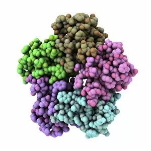
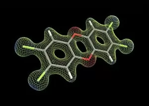
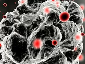
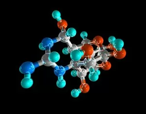
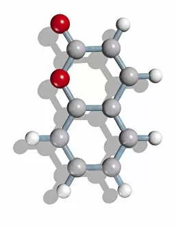
"Toxin: Unveiling the Dark Side of Nature's Chemistry" Aflatoxin, a silent threat lurking in our food, reveals its molecular model with intricate bonds and structures


All Professionally Made to Order for Quick Shipping
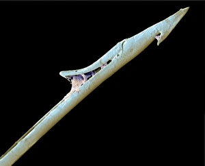
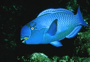
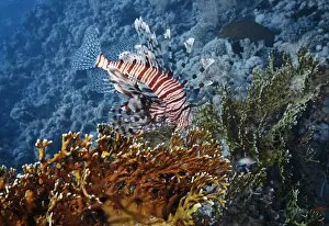
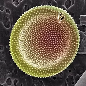
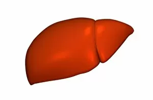
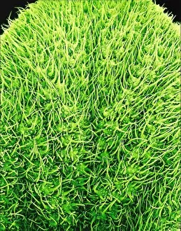
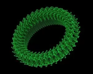
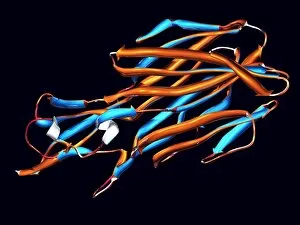
"Toxin: Unveiling the Dark Side of Nature's Chemistry" Aflatoxin, a silent threat lurking in our food, reveals its molecular model with intricate bonds and structures. In an artistic portrayal, Cholera bacteria showcases its deadly potential through vibrant artwork that captures the essence of this dangerous pathogen. The menacing Cholera toxin takes center stage as its molecular model unravels the secrets behind its ability to wreak havoc on human cells. Shiga toxin from E. Coli emerges as a sinister force, reminding us of the dangers posed by certain strains of this common bacterium. Hidden within the enchanting May apple, mandrake or wild lemon (Podophyllum peltatum), lies a potent toxin that serves as nature's defense mechanism against unsuspecting predators. The Penny bun or porcini mushroom (Boletus edulis) and its toxic counterpart, Suillellus luridus (Boletus perniciosus), stand side by side in their deceptive beauty – a reminder that not all mushrooms are safe for consumption. Ouida's litho artwork aptly titled "Toxin" portrays an eerie yet captivating representation of unseen dangers lurking beneath seemingly innocent surfaces. From the depths of rainforests to hidden corners in our homes, Pinktoe tarantulas and trapdoor spider nests serve as reminders that even arachnids possess venomous toxins for survival and defense. Strophanthus gratus unveils its lethal secret through delicate petals and thorny defenses – showcasing how plants can harness toxins to protect themselves from threats in their environment. The Poison arrow plant (Acokanthera oppositifolia) stands tall with vibrant blooms while concealing toxic compounds used historically by indigenous tribes for hunting purposes – illustrating nature's dual role as both healer and destroyer.