
Trematode Collection
Trematodes, also known as gastrointestinal nematodes, are a fascinating group of parasites that can wreak havoc on their hosts. Picture No


All Professionally Made to Order for Quick Shipping
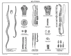
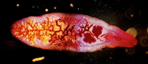
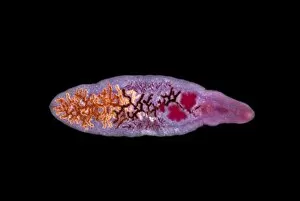
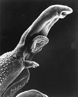
Trematodes, also known as gastrointestinal nematodes, are a fascinating group of parasites that can wreak havoc on their hosts. Picture No. 11675599 showcases the Lancet liver fluke C014 / 4846, one of the many species in this diverse family. Through scanning electron micrography, we get a closer look at the schistosome parasite - a notorious member of the trematode family. Research against Bilharzia has been crucial in understanding and combating these harmful organisms. Immature flukes have been captured under light micrographs, revealing their intricate structures and highlighting the need for effective treatment strategies. The Fluke larva is another intriguing subject seen through light microscopy; its unique features make it easily distinguishable from other parasites. In particular, cat flukes (C014 / 4858) have been studied extensively due to their impact on feline health. One captivating image depicts Schistosome flukes mating (micrograph C014 / 4867), shedding light on their reproductive behavior and life cycle intricacies. These microscopic creatures possess an astonishing ability to adapt and survive within various hosts. Lastly, we encounter the common liver fluke (C014 / 4847), which serves as a reminder of how prevalent these parasites can be worldwide. Through ongoing research efforts and advancements in parasitology, scientists strive to develop innovative solutions to combat trematode infections effectively. Understanding these complex organisms is vital for safeguarding human and animal health alike.