Fraud Blocker Skip to main content

Tumor Collection (page 3)

DDE-90026224, DDE-90026223, DDE-90026222, DDE-90026221, DDE-90026220, DDE-90026219

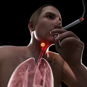
Background imageTumor Collection: Larynx cancer, artwork F007 / 6339

Larynx cancer, artwork F007 / 6339
Larynx cancer, computer artwork

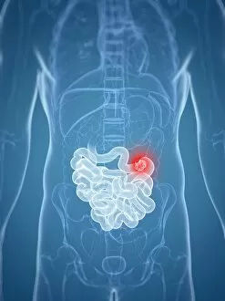
Background imageTumor Collection: Intestinal cancer, artwork F007 / 6664

Intestinal cancer, artwork F007 / 6664
Intestinal cancer, computer artwork

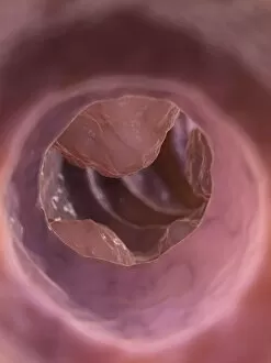
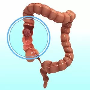
Background imageTumor Collection: Colon cancer, artwork F007 / 6862

Colon cancer, artwork F007 / 6862
Colon cancer, computer artwork

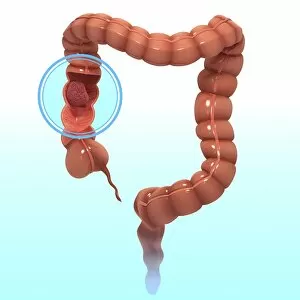
Background imageTumor Collection: Colon tumor, artwork F007 / 6861

Colon tumor, artwork F007 / 6861
Colon tumor, computer artwork

Background imageTumor Collection: Larynx cancer, artwork F007 / 6340

Larynx cancer, artwork F007 / 6340
Larynx cancer, computer artwork

Background imageTumor Collection: Larynx cancer, artwork F007 / 6337

Larynx cancer, artwork F007 / 6337
Larynx cancer, computer artwork

Background imageTumor Collection: Colon cancer, artwork F007 / 6876

Colon cancer, artwork F007 / 6876
Colon cancer, computer artwork

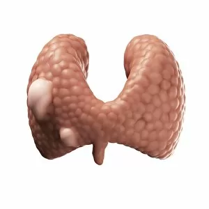
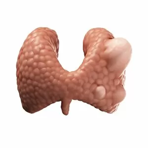
Background imageTumor Collection: Thyroid gland cancer, artwork F007 / 6128

Thyroid gland cancer, artwork F007 / 6128
Thyroid gland cancer. Computer artwork of follicular cancer of the thyroid gland
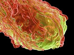
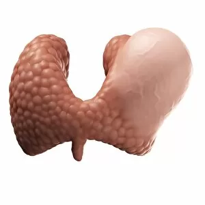
Background imageTumor Collection: Thyroid gland cancer, artwork F007 / 6181

Thyroid gland cancer, artwork F007 / 6181
Thyroid gland. Computer artwork of papillary cancer of the thyroid

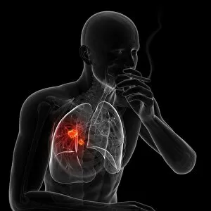
Background imageTumor Collection: Lung cancer due to smoking, artwork F007 / 6159

Lung cancer due to smoking, artwork F007 / 6159
Lung cancer due to smoking, computer artwork

Background imageTumor Collection: Lung cancer due to smoking, artwork F007 / 6161

Lung cancer due to smoking, artwork F007 / 6161
Lung cancer due to smoking, computer artwork

Background imageTumor Collection: Lung cancer due to smoking, artwork F007 / 6160

Lung cancer due to smoking, artwork F007 / 6160
Lung cancer due to smoking, computer artwork

Background imageTumor Collection: Thyroid gland cancer, artwork F007 / 6237

Thyroid gland cancer, artwork F007 / 6237
Thyroid gland cancer. Computer artwork of a solid adenocarcinoma of the thyroid

Background imageTumor Collection: Lung cancer due to smoking, artwork F007 / 6162

Lung cancer due to smoking, artwork F007 / 6162
Lung cancer due to smoking, computer artwork

Background imageTumor Collection: Colon cancer, artwork F008 / 0425

Colon cancer, artwork F008 / 0425
Colon cancer, computer artwork

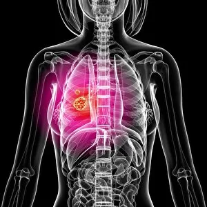
Background imageTumor Collection: Lung cancer, artwork F008 / 0116

Lung cancer, artwork F008 / 0116
Lung cancer, computer artwork

Background imageTumor Collection: Colon cancer, artwork F008 / 0585

Colon cancer, artwork F008 / 0585
Colon cancer, computer artwork

Background imageTumor Collection: Intestinal cancer, artwork F008 / 1199

Intestinal cancer, artwork F008 / 1199
Intestinal cancer, computer artwork

Background imageTumor Collection: Intestinal cancer, artwork F008 / 1197

Intestinal cancer, artwork F008 / 1197
Intestinal cancer, computer artwork

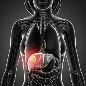
Background imageTumor Collection: Liver cancer, artwork F008 / 0127

Liver cancer, artwork F008 / 0127
Liver cancer, computer artwork

Background imageTumor Collection: Intestinal cancer, artwork F008 / 1198

Intestinal cancer, artwork F008 / 1198
Intestinal cancer, computer artwork

Background imageTumor Collection: Colon cancer, artwork F008 / 0678

Colon cancer, artwork F008 / 0678
Colon cancer, computer artwork

Background imageTumor Collection: Colon cancer, artwork F008 / 0672

Colon cancer, artwork F008 / 0672
Colon cancer, computer artwork

Background imageTumor Collection: Colon cancer, artwork F008 / 0306

Colon cancer, artwork F008 / 0306
Colon cancer, computer artwork

Background imageTumor Collection: Colon cancer, artwork F008 / 0050

Colon cancer, artwork F008 / 0050
Colon cancer, computer artwork

Background imageTumor Collection: Lung cancer, artwork F008 / 0214

Lung cancer, artwork F008 / 0214
Lung cancer, computer artwork

Background imageTumor Collection: Colon cancer, artwork F008 / 0424

Colon cancer, artwork F008 / 0424
Colon cancer, computer artwork

Background imageTumor Collection: Colon cancer, artwork F008 / 0519

Colon cancer, artwork F008 / 0519
Colon cancer, computer artwork

Background imageTumor Collection: Colon cancer, artwork F008 / 0142

Colon cancer, artwork F008 / 0142
Colon cancer, computer artwork

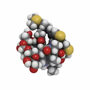
Background imageTumor Collection: Bleomycin drug molecule F005 / 6675

Bleomycin drug molecule F005 / 6675
Bleomycin, molecular model. Bleomycin is an antibiotic produced by the bacterium Streptomyces verticillus. It is used in the treatment of cancer and warts

Background imageTumor Collection: Methylparaben molecule

Methylparaben molecule
Molecular representation of methylparaben, one ester of parahydroxybenzoic acid. It is a commonly used preservative in cosmetics and pharmaceuticals

Background imageTumor Collection: Bleomycin drug molecule F005 / 6761

Bleomycin drug molecule F005 / 6761
Bleomycin, molecular model. Bleomycin is an antibiotic produced by the bacterium Streptomyces verticillus. It is used in the treatment of cancer and warts

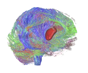
Background imageTumor Collection: Brain tumour, DTI MRI scan C017 / 7057

Brain tumour, DTI MRI scan C017 / 7057
Brain tumour. 3D diffusion tensor imaging (DTI) magnetic resonance imaging (MRI) scan of nerve pathways (coloured) in a brain with a tumour (red, centre right)

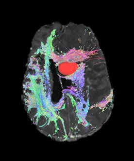
Background imageTumor Collection: Brain tumour, DTI MRI scan C017 / 7059

Brain tumour, DTI MRI scan C017 / 7059
Brain tumour. Axial diffusion tensor imaging (DTI) magnetic resonance imaging (MRI) scan of nerve pathways (coloured) in a brain with a tumour (red, upper centre)

Background imageTumor Collection: Brain tumour, DTI MRI scan C017 / 7058

Brain tumour, DTI MRI scan C017 / 7058
Brain tumour. 3D diffusion tensor imaging (DTI) magnetic resonance imaging (MRI) scan of nerve pathways (coloured) in a brain with a tumour (red, centre left)

Background imageTumor Collection: Tumour, computer artwork F007 / 0218

Tumour, computer artwork F007 / 0218
Tumour, computer artwork. Tumours are caused by the uncontrolled growth of previously normal cells. The resulting growth (green) can invade and damage surrounding tissue

Background imageTumor Collection: Tumour, computer artwork F007 / 0216

Tumour, computer artwork F007 / 0216
Tumour, computer artwork. Tumours are caused by the uncontrolled growth of previously normal cells. The resulting growth (blue) can invade and damage surrounding tissue

Background imageTumor Collection: Tumour, computer artwork F007 / 0214

Tumour, computer artwork F007 / 0214
Tumour, computer artwork. Tumours are caused by the uncontrolled growth of previously normal cells. The resulting growth (green) can invade and damage surrounding tissue

Background imageTumor Collection: Tumour, computer artwork F007 / 0212

Tumour, computer artwork F007 / 0212
Tumour, computer artwork. Tumours are caused by the uncontrolled growth of previously normal cells. The resulting growth (blue) can invade and damage surrounding tissue

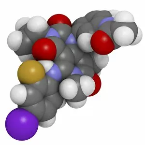
Background imageTumor Collection: Trametinib melanoma cancer drug F007 / 0197

Trametinib melanoma cancer drug F007 / 0197
Trametinib melanoma cancer drug, molecular model. Atoms are represented as spheres and are colour-coded: hydrogen (white), carbon (grey), oxygen (red), fluorine (dark yellow)

Background imageTumor Collection: Bone cancer, light micrograph F006 / 9810

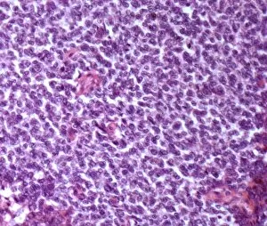
Bone cancer, light micrograph F006 / 9810
Bone cancer. Light micrograph of a section through an undifferentiated pleomorphic sarcoma of bone. This is a malignant (cancerous) tumour. Magnification: x200 when printed at 10 centimetres wide

Background imageTumor Collection: Immunotherapy, artwork

Immunotherapy, artwork
Immunotherapy. Computer artwork of immunotherapy being used to destroy a tumour (right). Immunotherapy uses the bodys immune system to fight a disease

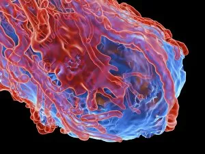
Background imageTumor Collection: Cancer cells spreading, artwork

Cancer cells spreading, artwork
Cancer cells spreading. Computer artwork showing cancerous cells (blue) migrating from a tumour (upper centre) into the blood stream (centre)

Background imageTumor Collection: Spinal cancer, MRI scan

Spinal cancer, MRI scan
Spinal cancer. Coloured magnetic resonance imaging (MRI) scan of a sagittal section through the cervical (neck) region of the spine of a 22-year-old patient

Background imageTumor Collection: Secondary spinal cancer, MRI scan

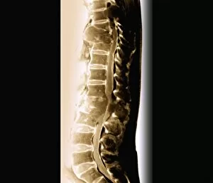
Secondary spinal cancer, MRI scan
Secondary spinal cancer. Magnetic resonance imaging (MRI) scan of a section through the spine of a 48-year-old patient, showing malignant (cancerous) tumours

Background imageTumor Collection: Metastatic cancer, artwork

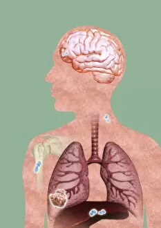
Metastatic cancer, artwork
Metastatic cancer. Computer artwork of a section through a persons upper body and head, showing a primary cancer in the right liver (lower left) that has spread (metastasised)

Background imageTumor Collection: Cancer blood supply, conceptual image C016 / 7505

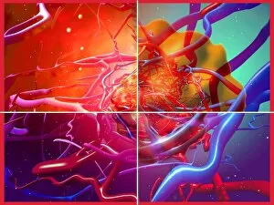
Cancer blood supply, conceptual image C016 / 7505
Cancer blood supply, conceptual image. Computer artwork representing the blood supply to a cancerous (malignant) tumour

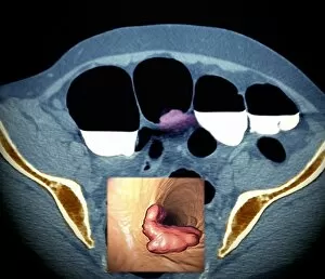
Background imageTumor Collection: Colon cancer, barium contrast CT scan

Colon cancer, barium contrast CT scan
Colon cancer. Coloured barium contrast computed tomography (CT) scan, and 3D CT colonoscopy image (inset), of an axial section through the sigmoid colon (large intestine) of a 54-year-old patient


All Professionally Made to Order for Quick Shipping

DDE-90026224, DDE-90026223, DDE-90026222, DDE-90026221, DDE-90026220, DDE-90026219, and DDE-90026218 - these are the codes that represent a battle fought by countless individuals against the silent enemy within: tumors. In 1889, Joseph Carey Merrick's haunting black and white photo captured not just his physical appearance but also the resilience of those facing unimaginable challenges. His story reminds us that behind every tumor lies a person with dreams and aspirations. An anterior view of female anatomy reveals breast tissue marred by a tumor. This image serves as a stark reminder of the importance of early detection and regular check-ups to safeguard our health. William Thomas Green Morton was an American pioneer in anesthesia who tragically lost his life at just 49 years old. His legacy lives on as medical professionals continue to explore innovative ways to combat tumors and improve patient outcomes. A medical illustration depicting a pilonidal cyst near the natal cleft serves as a visual representation of how even seemingly innocuous areas can be affected by tumors. It highlights the need for vigilance in monitoring our bodies for any signs or symptoms. Colon cancer is one of the most prevalent forms of this disease worldwide. A striking medical illustration showcases its different stages - from initial growth to advanced malignancy - urging us all to prioritize screenings and adopt healthy lifestyle choices. Tumors may be formidable foes, but they cannot extinguish hope or determination. Let us stand together in solidarity with those battling these insidious growths while supporting research efforts aimed at finding effective treatments and ultimately eradicating them altogether.