
Tumors Collection
"Tumors: Unveiling the Complexities of Abnormal Cell Growth" Secondary period syphilis symptoms on the body


All Professionally Made to Order for Quick Shipping
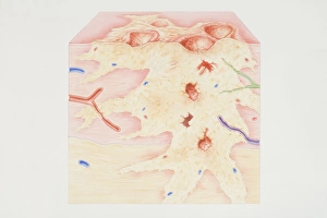
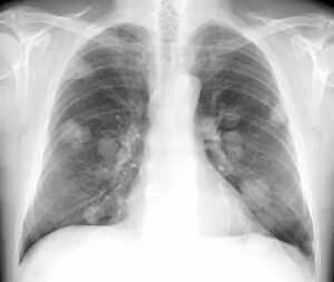
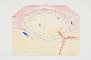
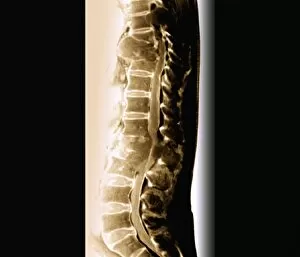
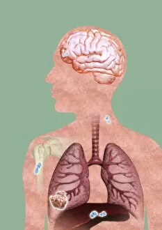
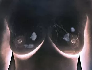
"Tumors: Unveiling the Complexities of Abnormal Cell Growth" Secondary period syphilis symptoms on the body: A closer look at how they are manifest as a result of secondary period syphilis, highlighting the importance of early detection and treatment. Cross-section diagram of a cancerous tumor: Explore the intricate components within a cancerous tumor, including calcium deposits, blood vessels, tumorous outgrowth, epithelial layer abnormalities, ulcerated areas, bleeding tendencies, nerve fiber involvement, dead tissue presence, and lymph vessel implications. Secondary lung cancers revealed through X-ray: Witness the impact of secondary lung cancers through an X-ray image that sheds light on their location and potential consequences for respiratory health. Cross-section diagram of a non-cancerous tumor: Delve into the structure of a non-cancerous tumor featuring a fibrous capsule enclosing tissues and accompanied by blood vessels—an intriguing glimpse into benign growths. Great Tit with head tumor in Warwickshire's waterside sanctuary: Marvel at nature's resilience as we encounter an adult Great Tit bird bravely facing life with a visible tumor growth on its head amidst the serene backdrop of Warwickshire's waterways in December. Cancer cells spreading depicted through artwork: Experience artistry capturing the relentless spread of cancer cells—a visual representation reminding us to remain vigilant against this formidable disease. 7 & 8 & Spinal cancer unravelled via MRI scans: Peer inside MRI scans revealing spinal cancers from various perspectives—each offering unique insights into this challenging condition affecting our central nervous system. 10 & Metastatic cancer portrayed through thought-provoking artwork.