Fraud Blocker Skip to main content

Ultrastructure Collection

Ultrastructure refers to the intricate

Background imageUltrastructure Collection: Mitochondrion, TEM

Mitochondrion, TEM
Mitochondrion. Coloured transmission electron micrograph (TEM) of mitochondria (green/blue) in heart muscle. Mitochondria are organelles found in the cytoplasm of eukaryotic cells

Background imageUltrastructure Collection: Mitochondria, TEM

Mitochondria, TEM
Mitochondria. Coloured transmission electron micrograph (TEM) of mitochondria (red) in heart muscle. Mitochondria are organelles found in the cytoplasm of eukaryotic cells

Background imageUltrastructure Collection: Heavy ion accelerator, Russia

Heavy ion accelerator, Russia
The IC-100 accelerator at the Flerov Laboratory of Nuclear Reactions of the Joint Institute for Nuclear Research in Russias Dubna

Background imageUltrastructure Collection: Influenza virus, artwork C016 / 8349

Influenza virus, artwork C016 / 8349
Influenza virus. Cut-away computer artwork of an influenza (flu) virus particle (virion). In each particles lipid envelope (green) are two types of protein spike, haemagglutinin (H)

Background imageUltrastructure Collection: Influenza virus, artwork C016 / 8348

Influenza virus, artwork C016 / 8348
Influenza virus. Cut-away computer artwork of an influenza (flu) virus particle (virion). In each particles lipid envelope (green) are two types of protein spike, haemagglutinin (H)

Background imageUltrastructure Collection: Influenza virus, artwork C016 / 8347

Influenza virus, artwork C016 / 8347
Influenza virus. Cut-away computer artwork of an influenza (flu) virus particle (virion). In each particles lipid envelope (green) are two types of protein spike, haemagglutinin (H)

Background imageUltrastructure Collection: Influenza virus, artwork C016 / 8346

Influenza virus, artwork C016 / 8346
Influenza virus. Cut-away computer artwork of an influenza (flu) virus particle (virion). In each particles lipid envelope (green) are two types of protein spike, haemagglutinin (H)

Background imageUltrastructure Collection: Influenza virus, artwork C016 / 8344

Influenza virus, artwork C016 / 8344
Influenza virus. Cut-away computer artwork of an influenza (flu) virus particle (virion). In each particles lipid envelope (green) are two types of protein spike, haemagglutinin (H)

Background imageUltrastructure Collection: Influenza virus, artwork C016 / 8345

Influenza virus, artwork C016 / 8345
This image may not be used in educational posters Influenza virus. Cut-away computer artwork of an influenza (flu) virus particle (virion)

Background imageUltrastructure Collection: Influenza virus, artwork C016 / 8342

Influenza virus, artwork C016 / 8342
Influenza virus. Computer artwork of an influenza (flu) virus particle (virion). In each particles lipid envelope (green) are two types of protein spike, haemagglutinin (H, red) and neuraminidase (N)

Background imageUltrastructure Collection: Influenza virus, artwork C016 / 8341

Influenza virus, artwork C016 / 8341
Influenza virus. Computer artwork of an influenza (flu) virus particle (virion). In each particles lipid envelope (green) are two types of protein spike, haemagglutinin (H, red) and neuraminidase (N)

Background imageUltrastructure Collection: Influenza virus, artwork C016 / 8340

Influenza virus, artwork C016 / 8340
Influenza virus. Computer artwork of an influenza (flu) virus particle (virion). In each particles lipid envelope (green) are two types of protein spike, haemagglutinin (H, red) and neuraminidase (N)

Background imageUltrastructure Collection: Influenza virus, artwork C018 / 2894

Influenza virus, artwork C018 / 2894
Influenza virus. Cut-away computer artwork of an influenza (flu) virus particle (virion). In each particles lipid envelope (blue) are two types of protein spike

Background imageUltrastructure Collection: Influenza virus, artwork C018 / 2893

Influenza virus, artwork C018 / 2893
Influenza virus. Cut-away computer artwork of an influenza (flu) virus particle (virion). In each particles lipid envelope (blue) are two types of protein spike

Background imageUltrastructure Collection: Influenza virus, artwork C018 / 2891

Influenza virus, artwork C018 / 2891
Influenza virus. Cut-away computer artwork of an influenza (flu) virus particle (virion). In each particles lipid envelope (blue) are two types of protein spike

Background imageUltrastructure Collection: Influenza virus, artwork C018 / 2890

Influenza virus, artwork C018 / 2890
Influenza virus. Cut-away computer artwork of an influenza (flu) virus particle (virion). In each particles lipid envelope (green) are two types of protein spike

Background imageUltrastructure Collection: Influenza virus, artwork C018 / 2892

Influenza virus, artwork C018 / 2892
Influenza virus. Cut-away computer artwork of an influenza (flu) virus particle (virion). In each particles lipid envelope (brown) are two types of protein spike

Background imageUltrastructure Collection: Petal papillae, SEM

Petal papillae, SEM
Petal papillae. Coloured scanning electron micrograph (SEM) of papillae on the surface of a cultivated primrose (Primula obconica) petal. Papillae are projections from epidermal cells

Background imageUltrastructure Collection: Artwork of the structure of a human hair

Artwork of the structure of a human hair

Background imageUltrastructure Collection: Illustration of the ultrastructure of a cell

Illustration of the ultrastructure of a cell
Illustration of the ultrastructure of a typical cell. The anatomical components of this cell are seen in three-dimensions and colour-coded


All Professionally Made to Order for Quick Shipping

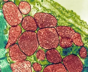
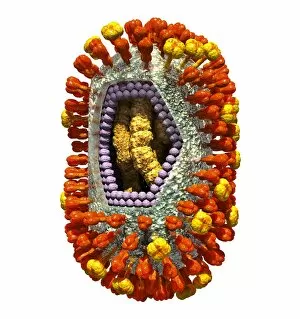
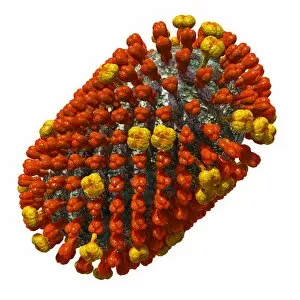
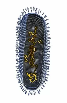
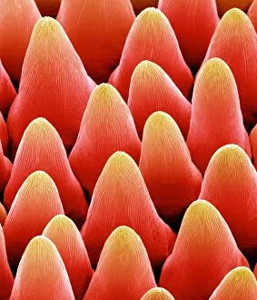
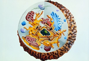
Ultrastructure refers to the intricate and detailed organization of cellular components that can only be observed using advanced imaging techniques such as transmission electron microscopy (TEM). One fascinating example is found within the mitochondrion, often referred to as the powerhouse of the cell. TEM images reveal its unique double membrane structure, with inner folds called cristae that increase surface area for energy production. In Russia, a remarkable scientific facility known as a heavy ion accelerator allows researchers to delve into the ultrastructural mysteries of various biological entities. Among these are captivating artworks depicting influenza viruses at different stages of their life cycle. Each artwork, such as C016 / 8349 or C016 / 8348, showcases the virus's outer envelope and internal components in exquisite detail. These mesmerizing TEM images provide valuable insights into how influenza viruses interact with host cells and cause infections. They unveil features like viral spikes on their surfaces that aid in attachment to target cells. By studying their ultrastructure, scientists gain crucial knowledge necessary for developing effective antiviral treatments and vaccines. The artwork series continues with C016 / 8347 through C016 / 8340, each revealing distinct aspects of influenza virus morphology and replication machinery. These visual masterpieces not only captivate viewers but also serve as invaluable tools for understanding viral pathogenesis. Through advancements in technology like TEM and access to cutting-edge facilities like heavy ion accelerators, scientists continue unraveling nature's hidden wonders at an unprecedented level of detail. The study opens up new avenues for research across various fields including cell biology, virology, and medicine – ultimately leading us closer to unlocking secrets that could revolutionize our understanding of life itself.