Fraud Blocker Skip to main content

Urethra Collection

Background imageUrethra Collection: Female urinary bladder, artwork

Female urinary bladder, artwork
Female urinary bladder, computer artwork. This organ receives and stores urine from the kidneys through the ureters (white tubes). The wrinkled bladder wall allows the bladder to expand if needed

Background imageUrethra Collection: Artwork of section through the female bladder

Artwork of section through the female bladder
Female bladder. Artwork showing a section through the female urinary system. The bladder is seen as a pink sac at centre. This is a hollow organ that holds urine

Background imageUrethra Collection: Digital cross section illustration of male reproductive system

Digital cross section illustration of male reproductive system

Background imageUrethra Collection: Microscopic view of Neisseria gonorrhoeae

Microscopic view of Neisseria gonorrhoeae

Background imageUrethra Collection: Anatomy of male urinary system

Anatomy of male urinary system

Background imageUrethra Collection: Early stage of kidney cancer with a tumor visible on the kidney

Early stage of kidney cancer with a tumor visible on the kidney

Background imageUrethra Collection: Male urinary system, artwork

Male urinary system, artwork
Male urinary system. Computer artwork of a male abdomen highlighting the urinary system. The two kidneys are at upper centre

Background imageUrethra Collection: Vandellia cirrhosa, candiru

Vandellia cirrhosa, candiru

Background imageUrethra Collection: Three dimensional view of female urinary system

Three dimensional view of female urinary system, profile view

Background imageUrethra Collection: Posterior view of urinary bladder showing seminal vesicles

Posterior view of urinary bladder showing seminal vesicles

Background imageUrethra Collection: Anatomy of female urinary system

Anatomy of female urinary system

Background imageUrethra Collection: Conceptual image of kidneys with ureter

Conceptual image of kidneys with ureter

Background imageUrethra Collection: Human urinary system

Human urinary system

Background imageUrethra Collection: Voiding cystourethrogram, X-rays C018 / 0577

Voiding cystourethrogram, X-rays C018 / 0577
Voiding cystourethrogram. X-ray of a 54 year old male patient undergoing a voiding cystourethrogram

Background imageUrethra Collection: Voiding cystourethrogram, X-rays C018 / 0578

Voiding cystourethrogram, X-rays C018 / 0578
Voiding cystourethrogram. Coloured X-ray of a 54 year old male patient undergoing a voiding cystourethrogram

Background imageUrethra Collection: Voiding cystourethrogram, X-rays C018 / 0579

Voiding cystourethrogram, X-rays C018 / 0579
Voiding cystourethrogram. Coloured X-ray of a 54 year old male patient undergoing a voiding cystourethrogram

Background imageUrethra Collection: Voiding cystourethrogram, X-rays C018 / 0465

Voiding cystourethrogram, X-rays C018 / 0465
Voiding cystourethrogram. Three coloured X-rays showing a 65 year old patient undergoing a voiding cystourethrogram

Background imageUrethra Collection: Bladder stones, artwork

Bladder stones, artwork
Bladder stones. Computer artwork showing a section through a human bladder containing bladder stones (whiteish, centre)


All products are expertly crafted, using premium materials, tailored to your specifications and promptly shipped


EDITORS COMMENTS

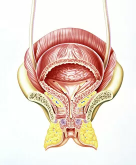
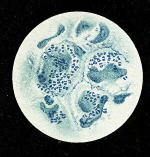
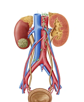
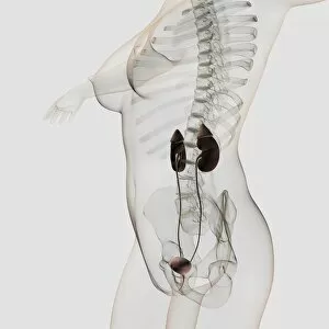
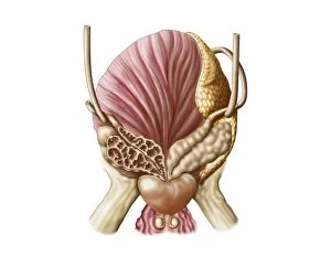
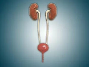
"Exploring the Intricacies of the Urethra: A Fascinating Journey Through Female Bladder Artwork" Delve into the captivating world of the urethra as we embark on a visual exploration through remarkable artwork. Witness an exquisite depiction of a section through the female bladder, revealing its intricate structure and function. Intriguingly, our journey takes us further as we encounter a lithograph showcasing a colony of Micrococcus Gonorrhoea found in the urethra back in 1906. This historical representation sheds light on early scientific discoveries surrounding this microorganism. Moving forward, we delve into cross-sectional anatomy with rectal exams conducted on normal males. These illustrations provide valuable insights into male reproductive systems and urinary tracts using both traditional and ultrasound technologies. Marvel at digital cross-section illustrations that unravel the complexity of male reproductive systems, offering an unprecedented glimpse into their inner workings. Additionally, microscopic views reveal Neisseria gonorrhoeae—a bacterium responsible for causing infections within this delicate organ. Surprisingly, our exploration extends beyond human anatomy to include intriguing aspects such as Vandellia cirrhosa or candiru—an infamous fish known for its peculiar parasitic behavior within urethras. Such unique encounters remind us of nature's diversity and adaptability. Lastly, let us not forget about kidney health; witness an early stage kidney cancer with a visible tumor on this vital organ. This serves as a reminder to prioritize regular check-ups and maintain awareness regarding potential risks associated with urological health. Through these captivating visuals encompassing both artistry and medical science, we gain profound appreciation for the intricacies housed within our urinary systems—highlighting their significance in maintaining overall well-being.

© Copyright Media Storehouse, All Rights Reserved 2003 - 2025
Facebook     Pinterest     100% Payment Secure     Reviews IO