Fraud Blocker Skip to main content

Urology Collection

"Exploring the Evolution of Urology: From Medieval Urine Wheels to Modern Innovations" Urology

Background imageUrology Collection: Medieval urine wheel

Medieval urine wheel. Labelled in Latin, this 15th century diagram shows some of the possible colours of urine (outer edge of circle) to help doctors diagnose urine-related diseases

Background imageUrology Collection: Kidney stone, 18th century

Kidney stone, 18th century

Background imageUrology Collection: ALBARRAN Y DOMINGEZ, Joaquin (1860 - 1912). Spanish urologist born in Cuba. (engraving)

ALBARRAN Y DOMINGEZ, Joaquin (1860 - 1912). Spanish urologist born in Cuba. (engraving)
CUL4940061 ALBARRAN Y DOMINGEZ, Joaquin (1860 - 1912). Spanish urologist born in Cuba. (engraving) by Unknown Artist; (add.info.: ALBARRAN Y DOMINGEZ, Joaquin (1860 - 1912)

Background imageUrology Collection: Brown-Beurger cystoscope, c. 1909

Brown-Beurger cystoscope, c. 1909
UCL273807 Brown-Beurger cystoscope, c.1909 by American School, (20th century); Science Collections, University College London

Background imageUrology Collection: Professor Felix Guyon, French surgeon and urologist, 1903

Professor Felix Guyon, French surgeon and urologist, 1903. Guyon (1831-1920) was the first professor of urology in Paris. He is also credited with discovering the ulnar canal in the wrist

Background imageUrology Collection: Doctor 17th Century Examines Examining Flask

Doctor 17th Century Examines Examining Flask
A 17th century European doctor examines a flask of urine from an elderly female patient. After a 17th century print by Quirin Boel after the painting by David Teniers

Background imageUrology Collection: Anatomy of male urinary system

Anatomy of male urinary system

Background imageUrology Collection: Early stage of kidney cancer with a tumor visible on the kidney

Early stage of kidney cancer with a tumor visible on the kidney

Background imageUrology Collection: Kidney stones, ultrasound scan C017 / 7381

Kidney stones, ultrasound scan C017 / 7381
Kidney stones. Ultrasound scan of a 83 year old female patient with two kidney stones (bright white) in the right kidney. Kidney stones, or calculi, are formed when mineral salts

Background imageUrology Collection: Male urinary system, artwork

Male urinary system, artwork
Male urinary system. Computer artwork of a male abdomen highlighting the urinary system. The two kidneys are at upper centre

Background imageUrology Collection: Bladder stones, ultrasound scan C017 / 7974

Bladder stones, ultrasound scan C017 / 7974
Bladder stones. Ultrasound scan of the bladder of a 58 year old male showing bladder stones (bright white). Bladder stones (cystoliths)

Background imageUrology Collection: Bladder epithelium, light micrograph

Bladder epithelium, light micrograph
Bladder epithelium. Light micrograph of a vertical section through the wall of the urinary bladder. The inner surface is at top

Background imageUrology Collection: Full bladder, X-ray cystography

Full bladder, X-ray cystography
Full bladder. X-ray cystogram of the pelvic region of a 50-year-old incontinent patient, showing urine (white) leaking (lower centre) from the bladder (round, centre) during a stress test

Background imageUrology Collection: ALBARRAN Y DOM͎GEZ, Joaqu�(1860 - 1912). Cuban

ALBARRAN Y DOM͎GEZ, Joaqu�(1860 - 1912). Cuban
ALBARRAN Y DOM͎ GEZ, Joaqu� (1860 - 1912). Cuban urologist. Engraving

Background imageUrology Collection: Anatomy of female urinary system

Anatomy of female urinary system

Background imageUrology Collection: Cross section illustration of human pelvis anatomy and male bladder

Cross section illustration of human pelvis anatomy and male bladder

Background imageUrology Collection: Human urinary system

Human urinary system

Background imageUrology Collection: Schematic of female organs in a funnel

Schematic of female organs in a funnel

Background imageUrology Collection: Richard Bright, engraved by H. Cock, from The National Portrait Gallery, Volume I

Richard Bright, engraved by H. Cock, from The National Portrait Gallery, Volume I
KW285310 Richard Bright, engraved by H. Cock, from The National Portrait Gallery, Volume I, published c.1820 (litho) by Say

Background imageUrology Collection: Testing strip being dipped into a urine sample

Testing strip being dipped into a urine sample

Background imageUrology Collection: Bladder stones, ultrasound scan C017 / 7262

Bladder stones, ultrasound scan C017 / 7262
Bladder stones. Ultrasound scan of the bladder of a 58 year old male showing bladder stones (bright white). Bladder stones (cystoliths)

Background imageUrology Collection: Bladder stones, ultrasound scan C017 / 7263

Bladder stones, ultrasound scan C017 / 7263
Bladder stones. Ultrasound scan of the bladder of a 58 year old male showing bladder stones (bright white). Bladder stones (cystoliths)

Background imageUrology Collection: Healthy adrenal glands, artwork

Healthy adrenal glands, artwork
Healthy adrenal glands, computer artwork

Background imageUrology Collection: Kidney stones, artwork C016 / 7531

Kidney stones, artwork C016 / 7531
Kidney stones. Computer artwork showing a section through a human kidney containing kidney stones (yellowish, right). The ureter (tube)

Background imageUrology Collection: Incontinence surgery, artwork C016 / 6843

Incontinence surgery, artwork C016 / 6843
Incontinence surgery. Artwork of the sacral region of a womans pelvis showing a schematic diagram for bladder and sacral surgery to treat incontinence

Background imageUrology Collection: Polycystic kidneys, CT scans C018 / 0571

Polycystic kidneys, CT scans C018 / 0571
Polycystic kidneys. Coloured computed tomography (CT) scans of the abdomen of a 28 year old patient with polycystic kidneys (pink)

Background imageUrology Collection: Polycystic kidneys, CT scans C018 / 0572

Polycystic kidneys, CT scans C018 / 0572
Polycystic kidneys. Coloured computed tomography (CT) scans of the abdomen of a 28 year old patient with polycystic kidneys (dark blue)

Background imageUrology Collection: Voiding cystourethrogram, X-rays C018 / 0577

Voiding cystourethrogram, X-rays C018 / 0577
Voiding cystourethrogram. X-ray of a 54 year old male patient undergoing a voiding cystourethrogram. This procedure involves liquid containing a radio-opaque dye being placed in the bladder via a

Background imageUrology Collection: Voiding cystourethrogram, X-rays C018 / 0578

Voiding cystourethrogram, X-rays C018 / 0578
Voiding cystourethrogram. Coloured X-ray of a 54 year old male patient undergoing a voiding cystourethrogram. This procedure involves liquid containing a radio-opaque dye being placed in the bladder

Background imageUrology Collection: Kidney infection, CT scan C018 / 0473

Kidney infection, CT scan C018 / 0473
Kidney infection. Coloured computed tomography (CT) scans of the abdomen of a 57 year old patient with a kidney infection

Background imageUrology Collection: Voiding cystourethrogram, X-rays C018 / 0579

Voiding cystourethrogram, X-rays C018 / 0579
Voiding cystourethrogram. Coloured X-ray of a 54 year old male patient undergoing a voiding cystourethrogram. This procedure involves liquid containing a radio-opaque dye being placed in the bladder

Background imageUrology Collection: Polycystic kidneys, CT scan C018 / 0573

Polycystic kidneys, CT scan C018 / 0573
Polycystic kidneys. Coloured computed tomography (CT) scan of the abdomen of a 28 year old patient with polycystic kidneys (dark blue)

Background imageUrology Collection: Voiding cystourethrogram, X-rays C018 / 0465

Voiding cystourethrogram, X-rays C018 / 0465
Voiding cystourethrogram. Three coloured X-rays showing a 65 year old patient undergoing a voiding cystourethrogram. This procedure involves liquid containing a radio-opaque dye being placed in

Background imageUrology Collection: Friedrich von Frerichs, German optician C018 / 7108

Friedrich von Frerichs, German optician C018 / 7108
Friedrich von Frerichs (1819-1885). 1889 engraving of the German ophthalmologist and pathologist Friedrich Theodor von Frerichs

Background imageUrology Collection: Bladder stones, ultrasound scan C017 / 7261

Bladder stones, ultrasound scan C017 / 7261
Bladder stones. Ultrasound scan of the bladder of a 58 year old male showing bladder stones (bright white). Bladder stones (cystoliths)

Background imageUrology Collection: E. coli bacteria, SEM C014 / 0385

E. coli bacteria, SEM C014 / 0385
E. coli bacteria. Coloured scanning electron micrograph (SEM) of Escherichia coli (E. coli) bacteria (rod-shaped) found in a urine sample from a patient with a urinary tract infection (UTI)

Background imageUrology Collection: E. coli bacteria, SEM C014 / 0386

E. coli bacteria, SEM C014 / 0386
E. coli bacteria. Coloured scanning electron micrograph (SEM) of Escherichia coli (E. coli) bacteria (rod-shaped) found in a urine sample from a patient with a urinary tract infection (UTI)

Background imageUrology Collection: Kidney stones, artwork

Kidney stones, artwork
Kidney stones. Computer artwork showing a section through a human kidney containing kidney stones (whiteish). The ureter (lower left)

Background imageUrology Collection: Bladder stones, artwork

Bladder stones, artwork
Bladder stones. Computer artwork showing a section through a human bladder containing bladder stones (whiteish, centre). The urethra can be seen (vertical, lower centre)


All Professionally Made to Order for Quick Shipping

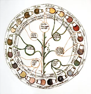
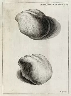
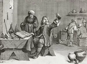
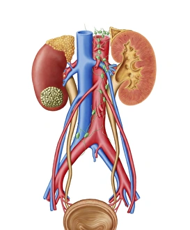
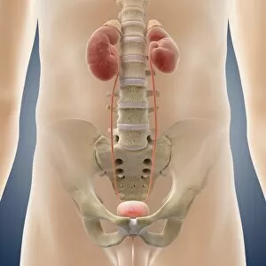
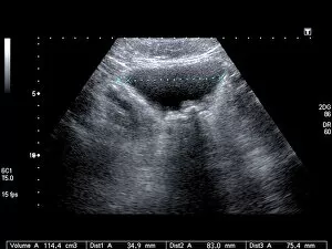
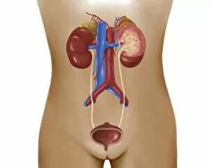
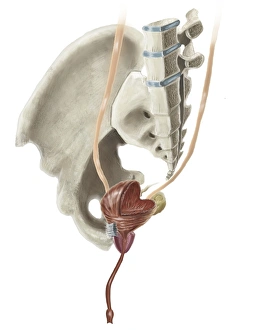
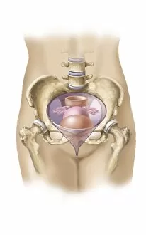
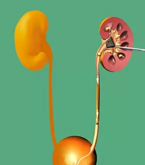
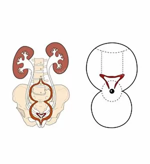
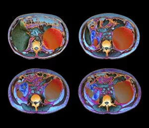
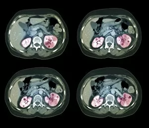
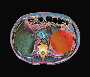
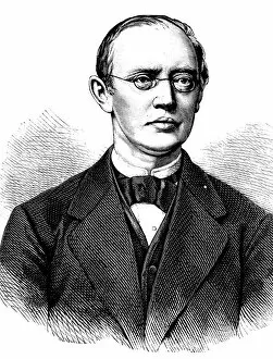
"Exploring the Evolution of Urology: From Medieval Urine Wheels to Modern Innovations" Urology, a medical specialty dedicated to the study and treatment of disorders related to the urinary system, has come a long way throughout history. In medieval times, physicians relied on peculiar methods like urine wheels to diagnose ailments. Fast forward to the 18th century when kidney stones plagued individuals, causing excruciating pain until innovative procedures were developed. One such advancement was the Brown-Beurger cystoscope in 1909, which revolutionized urological examinations by allowing direct visualization of the bladder and urethra. This breakthrough paved the way for remarkable surgeons like Professor Felix Guyon from France who made significant contributions in this field during his time. Joaquín Albarrán y Domínguez, a Cuban physician from 1860 - 1912, also played an essential role in urology's progress. His expertise helped shape modern techniques used today. Even back in the 17th century, doctors examined flasks as they sought ways to understand urinary diseases better. As technology advanced further into modern times, diagnostic tools improved significantly. For instance, X-ray cystography revealed full bladders with exceptional clarity while aiding in identifying early-stage kidney cancer tumors visible on kidneys themselves. Artwork depicting male urinary systems showcases intricate anatomical details that have been studied meticulously over centuries. Additionally, light micrographs capturing bladder epithelium provide invaluable insights into cellular structures within this vital organ. Today's urologists continue building upon these foundations laid by their predecessors through cutting-edge research and surgical interventions aimed at improving patients' lives worldwide. With each passing year comes new discoveries and advancements that push boundaries even further – all thanks to those who dedicated their lives to unraveling the mysteries of urology.