Fraud Blocker Skip to main content

Venous Collection (page 3)

"Exploring the Intricate Network Pathways

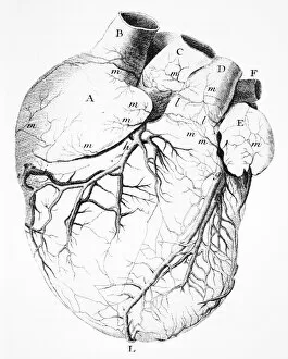
Background imageVenous Collection: Heart anatomy, 18th century

Heart anatomy, 18th century
Heart anatomy. 18th-century artwork showing the anatomy of the heart, including its coronary arteries. This artwork is from Traite de la structure du coeur, de son action

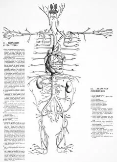
Background imageVenous Collection: Circulatory system, 16th century

Circulatory system, 16th century
Circulatory system. 16th-century artwork showing the heart and the arteries supplying blood to the body, as drawn by the Belgian anatomist Andreas Vesalius (1514-1564)

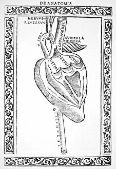
Background imageVenous Collection: Heart anatomy, 16th century

Heart anatomy, 16th century
Heart anatomy. 16th-century artwork of the anatomy of the heart, its blood vessels and nerves. This artwork is from Isagogae breves (1523)

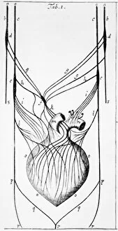
Background imageVenous Collection: Heart nerves, 17th century

Heart nerves, 17th century
Heart nerves, 17th-century artwork of the nerves that control the heart muscle and function. This artwork is from Tractatus de corde (1669) by the English physician Richard Lower (1631-1691)

Background imageVenous Collection: Venous system, 18th century

Venous system, 18th century. Artwork of the layout and appearance of the veins of the human circulatory system. This artwork is from Encyclopedie

Background imageVenous Collection: Circulatory system, 17th century

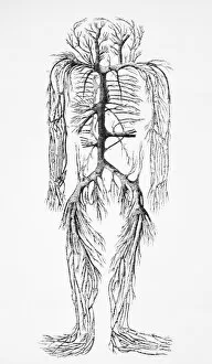
Circulatory system, 17th century
Circulatory system. Historical artwork of a human figure with internal organs and blood vessels shown. This manuscript is part of the collection gathered by the English antiquary Elias Ashmole

Background imageVenous Collection: Offering to the Greek god of medicine

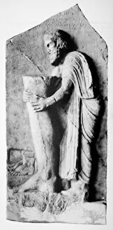
Offering to the Greek god of medicine. Ancient Greek sculpture of a man clasping a leg with visible veins. This was a votive offering to Asclepius, the Greek god of medicine

Background imageVenous Collection: Blood supply to alveoli, artwork

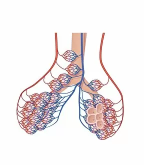
Blood supply to alveoli, artwork
Artwork showing the blood supply (arterial is red, venous is blue) transporting oxygen from and to the alveoli air sacs in the lungs

Background imageVenous Collection: Heart and lung anatomy, 17th century

Heart and lung anatomy, 17th century
Heart and lung anatomy. 17th-century artwork showing the anatomical structure of the heart and lungs. This artwork is from the posthumously published anatomical work De homine figuris (1662)

Background imageVenous Collection: Arm veins and valves, 16th century

Arm veins and valves, 16th century. Artwork of the discovery by the Italian anatomist and surgeon Hieronymus Fabricius (1533-1619) of valves within veins

Background imageVenous Collection: Small intestine structures, artwork

Small intestine structures, artwork
Small intestine structures. Artwork sequence of magnified views of the small intestine, with the magnification increasing from top to bottom

Background imageVenous Collection: Intestinal villi anatomy, artwork

Intestinal villi anatomy, artwork
Intestinal villi anatomy. Artwork showing six types of cell found on the surface of villi in the small intestine. Clockwise from upper right they are: enterocytes (green)

Background imageVenous Collection: Normal human heart, artwork

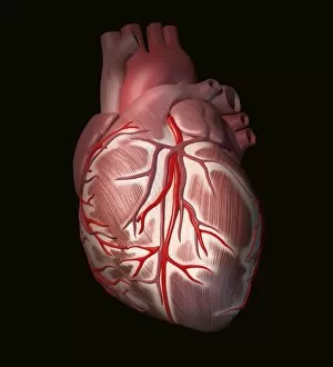
Normal human heart, artwork
Heart attack. Artwork showing the external appearance of a human heart as seen from the front during a myocardial infarction (heart attack)

Background imageVenous Collection: Blood vessels and kidneys, artwork

Blood vessels and kidneys, artwork
Blood vessels and kidneys. Computer artwork of a man with healthy blood vessels and kidneys. The kidneys are at lower centre

Background imageVenous Collection: Varicose veins, thermogram

Varicose veins, thermogram
MODEL RELEASED. Varicose veins. Varicose veins. Thermogram of the legs of a 56-year-old woman, showing severe varicose veins in the left leg (right)

Background imageVenous Collection: Female internal organs, artwork

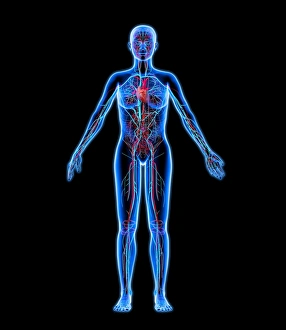
Female internal organs, artwork
Lymphatic system. Computer artwork of a naked woman with a healthy lymphatic system. Also seen are associated organs including the thymus gland (upper centre) and the spleen (centre right)

Background imageVenous Collection: Circulatory system and brain, artwork

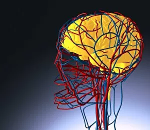
Circulatory system and brain, artwork
Circulatory system and brain. Computer artwork showing the blood vessels of the head and neck. Also seen is the brain (yellow)

Background imageVenous Collection: Cardiovascular system and brain, artwork

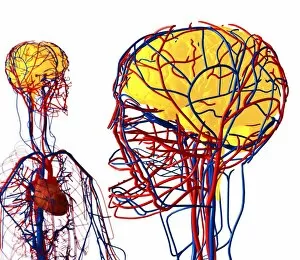
Cardiovascular system and brain, artwork
Cardiovascular system and brain. Computer artwork showing the blood vessels of the torso, arms, head and neck. Also seen is the brain (yellow)

Background imageVenous Collection: Cardiovascular system, artwork

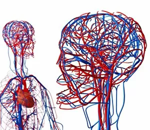
Cardiovascular system, artwork
Cardiovascular system. Computer artwork showing the blood vessels of the chest, arms, head and neck. The cardiovascular, or circulatory system

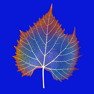
Background imageVenous Collection: Leaf veins

Leaf veins

Background imageVenous Collection: Leaf veins, X-ray

Leaf veins, X-ray
Leaf veins. Coloured X-ray of a leaf, exposing the venous structure. The main vein (midrib) runs down the centre of the leaf, branching off to form the other veins

Background imageVenous Collection: Diagram showing the circulation of the blood

Diagram showing the circulation of the blood
A diagram showing the circulation of the blood, with veins, arteries and capillaries


All Professionally Made to Order for Quick Shipping

"Exploring the Intricate Network Pathways: From Thyroid Gland Capillaries to Nephron Structures" Delve into the fascinating world anatomy with a captivating array of images and artwork. Witness the intricate web of thyroid gland capillaries, captured in stunning detail through scanning electron microscopy (SEM). Marvel at the complexity of thyroid gland blood vessels, also revealed by SEM, showcasing their vital role in hormone production. Embark on a journey through the cardiovascular system as depicted in exquisite artwork. Transport yourself back to the 16th century with a remarkable illustration depicting heart anatomy, showcasing how our understanding has evolved over centuries. Explore an engraving portraying the vascular system of the body, offering insight into its interconnectedness and importance for overall health. Discover the wonders of modern medical imaging technology as you peer into an MRI scan revealing the vascular system of the head. Gain a deeper appreciation for dental procedures with an artistic representation capturing dental filling polymerization - highlighting how even seemingly unrelated fields rely on proper venous circulation. Witness both beauty and pathology within this vast network. Admire an intricately crafted artwork illustrating thrombosed blood vessels - reminding us that maintaining healthy veins is crucial for optimal well-being. Immerse yourself in another masterpiece from Andreas Vesalius' De Humani Corporis Fabrica, published centuries ago but still revered today for its accurate depiction of human anatomy. From microscopic structures to grand engravings, these visual cues offer glimpses into our complex venous return system spanning across our upper torso and entire body. Appreciate every twist and turn as you navigate through this lifeline that ensures oxygen-rich blood reaches all corners efficiently. Intriguingly intertwined yet uniquely diverse, exploring "venous" reveals not only scientific marvels but also showcases art's ability to capture anatomical wonders throughout history – reminding us once again that knowledge truly knows no boundaries or limitations.