Fraud Blocker Skip to main content

Ventricles Collection

The intricate beauty of the human heart anatomy is captured in this captivating artwork

Background imageVentricles Collection: Human heart anatomy, artwork

Human heart anatomy, artwork
Human heart anatomy, cutaway computer artwork

Background imageVentricles Collection: Heart (Anon)

Heart (Anon)
the heart showing the ventricles, auricles, aorta, arteries, carotid branches and other associated vessels

Background imageVentricles Collection: Childs brain, MRI scan

Childs brain, MRI scan
Childs brain. Coloured magnetic resonance imaging (MRI) scan of an coronal (vertical) section through the head and brain of a healthy 3-year-old child, passing through the ears

Background imageVentricles Collection: Illustration depicting a Renaissance anatomical drawing (16th century; German)

Illustration depicting a Renaissance anatomical drawing (16th century; German)
5311488 Illustration depicting a Renaissance anatomical drawing (16th century; German. From Margarita Philosophica; 1503 by Reisch. Shows the ventricles and their relationship to the intellect)

Background imageVentricles Collection: Model of a brain (wax)

Model of a brain (wax)
BEN360191 Model of a brain (wax) by Susini, Clemente (1754-1814); Museo della Specola, Florence, Italy; (add.info.: cervello; scultura di cera; ); Photo eRaffaello Bencini; Italian, out of copyright

Background imageVentricles Collection: Model of a heart (wax)

Model of a heart (wax)
BEN360188 Model of a heart (wax) by Susini, Clemente (1754-1814); Museo della Specola, Florence, Italy; (add.info.: cuore; scultura di cera; ); Photo eRaffaello Bencini; Italian, out of copyright

Background imageVentricles Collection: Cross section of a normal heart

Cross section of a normal heart
Detail, Anatomy, Diagram, Muscle, Heart, Illustration, Biology, Chamber, Cardiac, Atrium, Cardiovascular, Cut Out, White Background, Vertical, Artwork, Cross Section, Color Image, Close Up

Background imageVentricles Collection: Brain fibres, DTI MRI scan C017 / 7036

Brain fibres, DTI MRI scan C017 / 7036
Brain fibres. 3D diffusion tensor imaging (DTI) magnetic resonance imaging (MRI) scan of a selection of nerve pathways (green/yellow) in the brain

Background imageVentricles Collection: Brain fibres, DTI MRI scan C017 / 7037

Brain fibres, DTI MRI scan C017 / 7037
Brain fibres. 3D diffusion tensor imaging (DTI) magnetic resonance imaging (MRI) scan of a selection of nerve pathways (green/yellow) in the brain

Background imageVentricles Collection: Brain fibres, DTI MRI scan C017 / 7038

Brain fibres, DTI MRI scan C017 / 7038
Brain fibres. 3D diffusion tensor imaging (DTI) magnetic resonance imaging (MRI) scan of a selection of nerve pathways (green/yellow) in the brain

Background imageVentricles Collection: Brain fibres, DTI MRI scan C017 / 7100

Brain fibres, DTI MRI scan C017 / 7100
Brain fibres. 3D diffusion tensor imaging (DTI) magnetic resonance imaging (MRI) scan of a selection of nerve pathways (blue, pink, green) in the brain. The front of the brain is at left

Background imageVentricles Collection: Heart chambers and sinus node, artwork C014 / 2030

Heart chambers and sinus node, artwork C014 / 2030
Heart chambers and sinus node (yellow). Cutaway artwork revealing the four internal heart chambers and the heart valves and tendons (white). The two upper chambers are the atria

Background imageVentricles Collection: Heart chamber anatomy, artwork C014 / 2029

Heart chamber anatomy, artwork C014 / 2029
Heart chamber anatomy. Cutaway artwork revealing the four internal heart chambers and the heart valves and tendons (white). The two upper chambers are the atria

Background imageVentricles Collection: Human head, MRI and 3D CT scans C016 / 6396

Human head, MRI and 3D CT scans C016 / 6396
Human head. Coloured composite image of a magnetic resonance imaging (MRI) scan of the brain and a 3D computed tomography (CT) scan of the skull of a 35 year old patient

Background imageVentricles Collection: Human head, MRI and 3D CT scans C016 / 6398

Human head, MRI and 3D CT scans C016 / 6398
Human head. Coloured composite image of a magnetic resonance imaging (MRI) scan of the brain and a 3D computed tomography (CT) scan of the skull of a 35 year old patient

Background imageVentricles Collection: Hydrocephalus, CT scan C018 / 0568

Hydrocephalus, CT scan C018 / 0568
Hydrocephalus. Coloured computed tomography (CT) scan of the brain of a 22 year old patient with hydrocephalus (fluid in the brain)

Background imageVentricles Collection: Heart, illustration C018 / 0797

Heart, illustration C018 / 0797
Heart, artwork. The heart is a hollow muscle that pumps blood around the body. Deoxygenated blood from the body is returned to the hearts right atrium (chamber)

Background imageVentricles Collection: Hydrocephalus, CT scan C018 / 0567

Hydrocephalus, CT scan C018 / 0567
Hydrocephalus. Coloured computed tomography (CT) scan of the brain of a 22 year old patient with hydrocephalus (fluid in the brain)

Background imageVentricles Collection: Hydrocephalus, CT scans C018 / 0566

Hydrocephalus, CT scans C018 / 0566
Hydrocephalus. Coloured computed tomography (CT) scans of the brain of a 22 year old patient with hydrocephalus (fluid in the brain)

Background imageVentricles Collection: Human heart, historical anatomical model

Human heart, historical anatomical model
Human heart. 1950s model showing the anatomy of the human heart

Background imageVentricles Collection: Hearts coronary blood vessels, artwork

Hearts coronary blood vessels, artwork
Hearts coronary blood vessels. Artwork of a human heart seen from the front, sectioned to show the internal chambers and the structure of the hearts major blood vessels (across top)

Background imageVentricles Collection: Heart ventricle anatomy, artwork C016 / 2875

Heart ventricle anatomy, artwork C016 / 2875
Ventricular diastole. Artwork showing a section through a human heart and its associated blood vessels during ventricular diastole. The heart is composed of four chambers

Background imageVentricles Collection: Choroid plexus secretory cells, SEM

Choroid plexus secretory cells, SEM
Choroid plexus secretory cells. Coloured scanning electron micrograph (SEM) of a freeze-fractured choroid plexus from a brain

Background imageVentricles Collection: Alzheimers disease brain, coloured CT scan

Alzheimers disease brain, coloured CT scan
Alzheimers disease brain, coloured computed tomography (CT) scan. This is the brain of an 81 year old woman. The front of the brain is at top

Background imageVentricles Collection: Neurogenesis, artwork

Neurogenesis, artwork
Neurogenesis. Artwork of an adult brain, revealing neurogenesis (nerve cell synthesis) sites. It was once believed that adult brains could not synthesise new neurons (nerve cells)

Background imageVentricles Collection: Contour map of the heart

Contour map of the heart, computer artwork. The contours on this map describe the surface shape of the heart, shown here from the front

Background imageVentricles Collection: Electrical conduction in the heart, artwork

Electrical conduction in the heart, artwork. Each heartbeat is initiated by an electrical impulse from the sinoatrial (SA) node (green oval) in the right atrium (upper heart chamber)

Background imageVentricles Collection: Dissected heart, light micrograph

Dissected heart, light micrograph
Dissected heart. Light micrograph of a section through a whole heart, showing the chambers and some blood. The heart is a hollow muscular organ that pumps blood around the body

Background imageVentricles Collection: Heart structure

Heart structure
Artwork of the internal structure of the heart. On the left, the right atrium (top cavity) and right ventriculum (bottom) receive deoxygenated blood from the body through the superior

Background imageVentricles Collection: Atrial fibrillation

Atrial fibrillation. Artwork of a section through a human heart during atrial fibrillation, a rapid, irregular heartbeat. In the background are ECG (electrocardiogram) traces

Background imageVentricles Collection: Internal heart anatomy, artwork

Internal heart anatomy, artwork
Internal heart anatomy. Cutaway artwork revealing the four internal heart chambers and the heart valves and tendons (white). The two upper chambers are the atria

Background imageVentricles Collection: Brain lining, SEM

Brain lining, SEM
Brain lining. Coloured scanning electron micrograph (SEM) of the lining of the brain, showing the ciliary hairs (green) of ependymal cells

Background imageVentricles Collection: Anatomy of the heart, artwork

Anatomy of the heart, artwork
Artwork of a healthy human heart showing the main blood vessels (at top) that service it and the internal chambers (atria and ventricles) with cardiac valves (blue)

Background imageVentricles Collection: Heart valves, artwork

Heart valves, artwork
Heart valves. Cutaway artwork of the inside of the heart, showing the various valves. The first two artworks (left and centre) show the atrioventricular valves between the hearts atria (top)

Background imageVentricles Collection: Dogfish head, transverse section

Dogfish head, transverse section
Dogfish head. Light micrograph of a transverse section through the head of a lesser spotted dogfish (Scyliorhinus canicula), in the region of the eyes (upper left and upper right)


All Professionally Made to Order for Quick Shipping

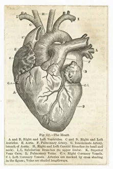
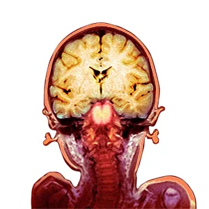
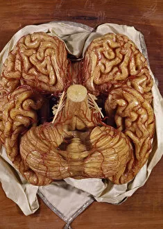
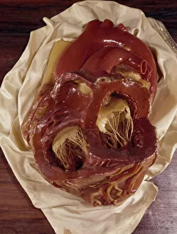
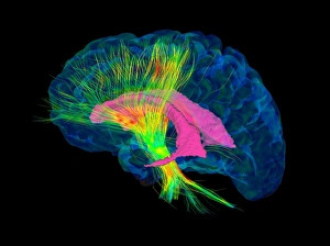
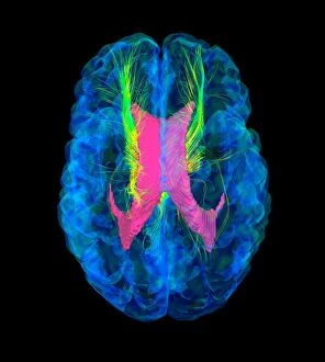
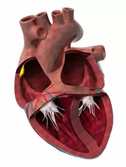
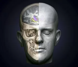
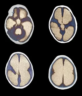
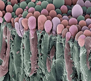
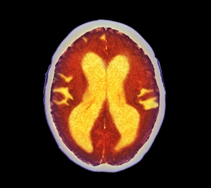
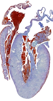
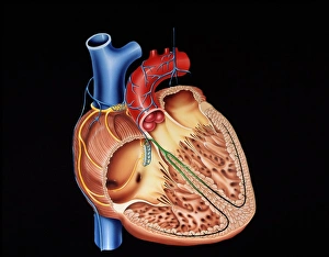
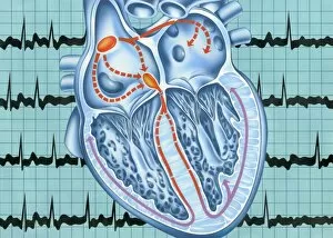
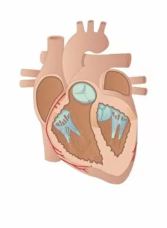
The intricate beauty of the human heart anatomy is captured in this captivating artwork, showcasing the ventricles that play a vital role in pumping blood throughout our bodies. In an anonymous masterpiece titled "Heart, " we witness the complexity and elegance of this organ, with its cross-section revealing the chambers known as ventricles. Moving from hearts to brains, a child's MRI scan unveils not only their developing mind but also provides glimpses into the interconnectedness between these two essential organs. Sections through the brain showcase both cerebellum and ventricles, highlighting their significance in maintaining cognitive functions. Delving deeper into microscopic details, an SEM image showcases brain lining while another MRI scan of a child's brain reveals astonishing intricacies. These images remind us of how delicate yet resilient our brains are. Venturing beyond traditional anatomical structures, we encounter fallopian tube cells under SEM examination. Although seemingly unrelated to ventricles at first glance, it serves as a reminder that every part of our body plays a unique role in maintaining overall health and balance. Stepping into wax models brings forth tangible representations of both brains and hearts. A model brain stands as a testament to scientific advancements allowing us to understand complex neural networks within those precious ventricles. Similarly, a model heart exhibits its own set of chambers including those crucial ventricles responsible for circulation. A cross-section image further emphasizes normal heart anatomy with prominently displayed ventricles working harmoniously alongside other components like valves and arteries. Ultimately, these various depictions highlight the importance and wonder surrounding our intricate biology – from macroscopic views encompassing entire organs down to microscopic examinations uncovering cellular wonders within them. The mesmerizing presence reminds us that life pulsates through each beat they orchestrate within our chests – an awe-inspiring symphony keeping us alive day by day.