Fraud Blocker Skip to main content

Ventricular Collection

"Exploring the Intricacies of Ventricular Function

Background imageVentricular Collection: ECGs of a normal heart rate, artwork

ECGs of a normal heart rate, artwork
Electrocardiograms (ECG) of a normal heart rate, artwork. An ECG measures the electrical activity of the heart. The wave on the ECG appears upwards or downwards depending upon whether the electrical

Background imageVentricular Collection: Irregular heartbeat

Irregular heartbeat. Conceptual image of a chest X-ray overlaid with an electrocardiogram (heart monitor) trace showing extrasystole

Background imageVentricular Collection: Ventricular extrasystole, anatomical artw

Ventricular extrasystole, anatomical artw
Atrial extrasystole. Anatomical computer artwork of a transverse section through a human heart during ventricular contraction in extrasystole

Background imageVentricular Collection: Extrasystole heartbeats, ECG artwork

Extrasystole heartbeats, ECG artwork. Diagram showing the electrical activity of the atrium (top) and ventricle (bottom) of a human heart during extrasystole

Background imageVentricular Collection: Ventricular systole, anatomical artwork

Ventricular systole, anatomical artwork
Ventricular systole. Anatomical computer artwork of a transverse section through a healthy human heart during ventricular systole of the cardiac cycle (heartbeat)

Background imageVentricular Collection: Hearts coronary blood vessels, artwork

Hearts coronary blood vessels, artwork
Hearts coronary blood vessels. Artwork of a human heart seen from the front, sectioned to show the internal chambers and the structure of the hearts major blood vessels (across top)

Background imageVentricular Collection: Heart ventricle anatomy, artwork C016 / 2875

Heart ventricle anatomy, artwork C016 / 2875
Ventricular diastole. Artwork showing a section through a human heart and its associated blood vessels during ventricular diastole. The heart is composed of four chambers

Background imageVentricular Collection: Alzheimers disease brain, coloured CT scan

Alzheimers disease brain, coloured CT scan
Alzheimers disease brain, coloured computed tomography (CT) scan. This is the brain of an 81 year old woman. The front of the brain is at top

Background imageVentricular Collection: Illustration of section through human heart

Illustration of section through human heart
Illustration of a longitudinal section through the human heart in hypertrophic cardiomyopathy, showing massive enlargement of the muscular septum separating the left and right ventricles

Background imageVentricular Collection: Artwork of heart in congestive heart failure

Artwork of heart in congestive heart failure
Heart failure. Cutaway illustration of a human heart showing congestive ventricular heart failure. Blood vessels connecting the heart to the rest of the body are at top

Background imageVentricular Collection: LM of section whole heart showing LV infarct

LM of section whole heart showing LV infarct
Infarcted left ventricle: light micrograph of a section through the ventricles of a whole human heart, showing an area of infarction (muscle necrosis or death)

Background imageVentricular Collection: Artwork of heart & ECG trace in heart failure

Artwork of heart & ECG trace in heart failure
Heart failure. Cutaway illustration of a human heart and an ECG (electrocardiogram) trace in left ventricular heart failure

Background imageVentricular Collection: Coloured chest X-ray of ventricular cardiomyopathy

Coloured chest X-ray of ventricular cardiomyopathy

Background imageVentricular Collection: Neurogenesis, artwork

Neurogenesis, artwork
Neurogenesis. Artwork of an adult brain, revealing neurogenesis (nerve cell synthesis) sites. It was once believed that adult brains could not synthesise new neurons (nerve cells)

Background imageVentricular Collection: ECG of a normal heart rate, artwork

ECG of a normal heart rate, artwork
Electrocardiogram (ECG) of a normal heart rate, artwork. An ECG measures the electrical activity of the heart. Contractions are caused by electrical signals between the upper (atrial)

Background imageVentricular Collection: Trabeculae carneae in the heart, SEM

Trabeculae carneae in the heart, SEM
Trabeculae carneae in the left ventricle of the heart, coloured scanning electron micrograph (SEM). Endocardial connective tissue is also seen


All Professionally Made to Order for Quick Shipping

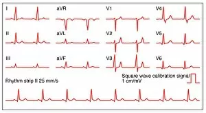
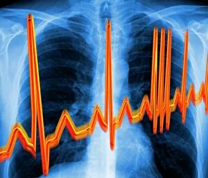
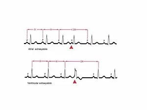
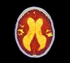
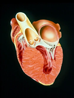
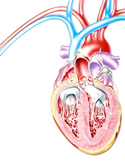
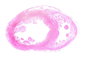
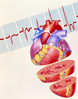
"Exploring the Intricacies of Ventricular Function: From ECGs to Artwork" Delve into the fascinating world function as we unravel its mysteries through a captivating blend of scientific visuals and artistic interpretations. Starting with ECGs showcasing a normal heart rate, witness the rhythmic beauty that lies within our chests. But what happens when irregularities arise? Brace yourself for an exploration of irregular heartbeat patterns, revealing the complexities hidden beneath each beat. Venturing deeper, we take you on a journey through sections of the cerebellum and ventricular system. Marvel at how these intricate structures work in harmony to maintain equilibrium within our bodies. Through anatomical artwork, get up close and personal with the human heart - an organ so vital yet enigmatic. Discover its inner workings as we unveil detailed depictions extrasystole, shedding light on those occasional abnormal heartbeats that can leave us breathless. ECG artwork brings further clarity to these extrasystolic episodes, allowing us to visualize their impact on our cardiac rhythm. Witness how even slight deviations from normality can have profound effects on our overall health. Returning to anatomical artwork once more, immerse yourself in the wonders systole - a crucial phase where blood is efficiently pumped out into circulation. Appreciate this synchronized dance between muscle fibers as they contract and relax with precision. But let's not forget about one essential aspect – coronary blood vessels. Admire stunning artworks depicting these lifelines that nourish our hearts day in and day out. Understand their significance in maintaining optimal cardiac function while marveling at their intricate branching patterns. Intriguingly complex yet undeniably beautiful, explore every nook and cranny of heart ventricle anatomy through meticulously crafted artwork C016 / 2875. Uncover secrets held by this remarkable structure that keeps us alive every single moment.