
Venule Collection
Exploring the intricate world of venules: from thyroid gland capillaries to kidney blood vessels, SEM images reveal their fascinating structures


All Professionally Made to Order for Quick Shipping
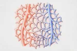
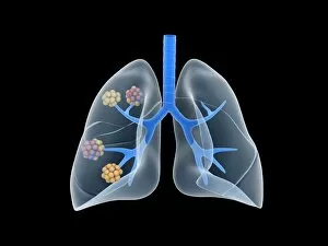
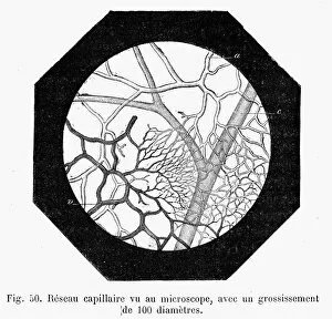
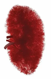
Exploring the intricate world of venules: from thyroid gland capillaries to kidney blood vessels, SEM images reveal their fascinating structures. Witness the delicate network within the thyroid gland and marvel at the complexity of its capillaries under a scanning electron microscope. Journey into the kidneys as SEM unveils the beauty of blood vessels and glomeruli, showcasing their vital role in filtration. Delve deeper into medical science as we explore diseased alveoli in the lung, reminding us of the importance of healthy venules for proper respiratory function. A diagram illustrates how these tiny vessels connect arterioles and venules, highlighting their crucial role in maintaining circulation throughout our bodies. Conceptual images transport us to an ethereal realm where alveoli come alive with vibrant colors, emphasizing their significance in gas exchange within our lungs. Let's appreciate these captivating venules that weave together various organs and systems, ensuring our well-being on a microscopic level.