Fraud Blocker Skip to main content

Vesicle Collection (#2)

"Exploring the Intricate World of Vesicles: Unveiling Cellular Secrets" Synapse nerve junction

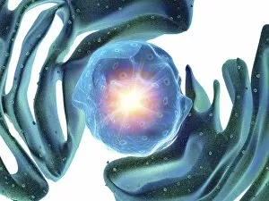
Background imageVesicle Collection: Nucleus and endoplasmic reticulum C015 / 6782

Nucleus and endoplasmic reticulum C015 / 6782
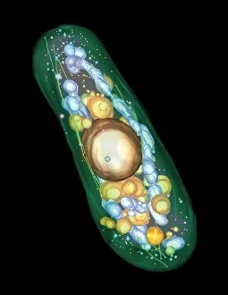
Computer artwork showing part of a human or eukaryotic cell. In the middle the nucleus which has a membrane with nuclear pores. Inside the nucleus is the DNA
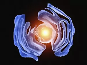
Background imageVesicle Collection: Nucleus and endoplasmic reticulum C015 / 6781

Nucleus and endoplasmic reticulum C015 / 6781
Computer artwork showing part of a human or eukaryotic cell. In the middle the nucleus which has a membrane with nuclear pores. Inside the nucleus is the DNA

Background imageVesicle Collection: Animal cell organelles, artwork C015 / 6775

Animal cell organelles, artwork C015 / 6775
Animal cell organelles. Computer artwork showing the organelles in a eukaryotic cell. This is an animal cell. Structures include the nucleus (centre) which has a membrane with nuclear pores

Background imageVesicle Collection: Animal cell organelles, artwork C015 / 6778

Animal cell organelles, artwork C015 / 6778
Animal cell organelles. Computer artwork showing the organelles in a eukaryotic cell. This is an animal cell. Structures include the nucleus (centre) which has a membrane with nuclear pores

Background imageVesicle Collection: Animal cell organelles, artwork C015 / 6777

Animal cell organelles, artwork C015 / 6777
Animal cell organelles. Computer artwork showing the organelles in a eukaryotic cell. This is an animal cell. Structures include the nucleus (centre) which has a membrane with nuclear pores

Background imageVesicle Collection: Nucleus and endoplasmic reticulum C015 / 6772

Nucleus and endoplasmic reticulum C015 / 6772
Computer artwork showing part of a human or eukaryotic cell. In the middle the nucleus which has a membrane with nuclear pores. Inside the nucleus is the DNA

Background imageVesicle Collection: Nucleus and endoplasmic reticulum C015 / 6768

Nucleus and endoplasmic reticulum C015 / 6768
Computer artwork showing part of a human or eukaryotic cell. In the middle the nucleus which has a membrane with nuclear pores. Inside the nucleus is the DNA

Background imageVesicle Collection: Nucleus and endoplasmic reticulum C015 / 6767

Nucleus and endoplasmic reticulum C015 / 6767
Computer artwork showing part of a human or eukaryotic cell. In the middle the nucleus which has a membrane with nuclear pores. Inside the nucleus is the DNA

Background imageVesicle Collection: Golgi apparatus, artwork C015 / 6764

Golgi apparatus, artwork C015 / 6764
Computer artwork of the Golgi apparatus of the human cell. This organelle functions as a central delivery system for the cell

Background imageVesicle Collection: HeLa cell dying, SEM C017 / 8304

HeLa cell dying, SEM C017 / 8304
HeLa cell dying, coloured scanning electron micrograph (SEM). The cell in the foreground is undergoing apoptosis, or programmed cell death. Apoptosis occurs when a cell becomes old or damaged

Background imageVesicle Collection: Intracellular transport, artwork C013 / 5001

Intracellular transport, artwork C013 / 5001
Intracellular transport. Computer artwork of a vesicle (sphere) being transported along a microtubule (blue and green) by a kinesin motor protein (orange)

Background imageVesicle Collection: Intracellular transport, artwork C013 / 4997

Intracellular transport, artwork C013 / 4997
Intracellular transport. Computer artwork of vesicles (spheres) being transported from a Golgi body (blue, left) around the cell by microtubules (string-like)

Background imageVesicle Collection: Intracellular transport, artwork C013 / 4995

Intracellular transport, artwork C013 / 4995
Intracellular transport. Computer artwork of vesicles (spheres) being transported from a Golgi body (blue, top) around the cell by microtubules (string-like)

Background imageVesicle Collection: Pancreatic cell, TEM

Pancreatic cell, TEM
Pancreatic cell. Coloured transmission electron micrograph (TEM) of part of an acinar (exocrine) pancreatic cell. Mitochondria (red) are seen in the cells endoplasmic reticulum (yellow)

Background imageVesicle Collection: Pancreas cells, SEM

Pancreas cells, SEM
Pancreas cells. Coloured scanning electron micrograph (SEM) of acinar (exocrine) pancreatic cells. Acinar cells produce and excrete digestive enzymes to the small intestine, via the pancreatic ducts

Background imageVesicle Collection: Alveolar cell, TEM

Alveolar cell, TEM
Alveolar cell. Coloured transmission electron micrograph (TEM) of a section through a type II alveolar cell. This cell is found in the alveoli (air sacs) of the lungs

Background imageVesicle Collection: Astrocyte / neuron synapse in the brain

Astrocyte / neuron synapse in the brain
Astrocyte/neuron synapse in the brain. Artwork of a nerve cell (neuron, yellow) synapse in the brain with an associated regulatory astrocyte cell (red)

Background imageVesicle Collection: Skin lesions

Skin lesions. Artwork from Robert Willans 1808 medical textbook, On Cutaneous Diseases, showing 16 different kinds of disorders affecting the skin

Background imageVesicle Collection: Simulated space ice

Simulated space ice. Light micrograph of vesicles (round structures) in ice formed under conditions simulating those in space

Background imageVesicle Collection: Inside of an imaginary cell

Inside of an imaginary cell
Inside a cell. Panoramic view from inside an imaginary cell. The densely folded endoplasmic reticulum, a membrane that divides the cell into compartments, is shown in yellow and red

Background imageVesicle Collection: Liposomes, SEM

Liposomes, SEM

Background imageVesicle Collection: Evolving protocell, artwork

Evolving protocell, artwork
Evolving protocell. Image 3 of 5. Artwork showing a protocell (artificial cell). This protocell was formed from a synthetic primeval soup containing PNA (peptide nucleic acid, orange)

Background imageVesicle Collection: Computer enhanced coloured LM of lipsomes

Computer enhanced coloured LM of lipsomes
Liposomes. Computer enhanced coloured light micrograph of liposomes. A liposome is an artificial vesicle made from phospholipids which can have many uses in biology

Background imageVesicle Collection: Protocell proliferation, artwork

Protocell proliferation, artwork
Protocell proliferation. Image 5 of 5. Artwork showing a protocell (artificial cell) dividing to produce two daughter cells

Background imageVesicle Collection: Smell receptors, TEM

Smell receptors, TEM
Smell receptors. Coloured transmission electron micrograph (TEM) of a section through smell receptors (cilia) projecting from an olfactory neurone (blue)

Background imageVesicle Collection: Liposomes, TEM

Liposomes, TEM

Background imageVesicle Collection: Pancreas cell, SEM

Pancreas cell, SEM
Pancreas cell. Coloured scanning electron micrograph (SEM) of an acinar (exocrine) pancreatic cell. Acinar cells produce and excrete digestive enzymes to the small intestine

Background imageVesicle Collection: Newly formed protocell, artwork

Newly formed protocell, artwork
Newly formed protocell. Image 2 of 5. Artwork showing cellular components inside a basic cell membrane. This protocell (artificial cell)

Background imageVesicle Collection: Energised protocell, artwork

Energised protocell, artwork
Energised protocell. Image 4 of 5. Artwork showing light photons energising a protocell (artificial cell). This protocell was formed from a synthetic primeval soup containing PNA

Background imageVesicle Collection: Basophil white blood cells, artwork

Basophil white blood cells, artwork
Basophil white blood cells. Computer artwork of basophil white blood cells, showing their lobed nuclei (red). Basophils are the smallest and least common of the white blood cells

Background imageVesicle Collection: Cholesterol particle, artwork

Cholesterol particle, artwork. This is low density lipoprotein (LDL), or bad, cholesterol. It consists of a core of esterified cholesterol molecules(green and blue)

Background imageVesicle Collection: Familial hypercholesterolaemia, diagram

Familial hypercholesterolaemia, diagram
Familial hypercholesterolaemia. Diagram showing the biochemical processes that take place in a case of the genetic disorder familial hypercholesterolaemia

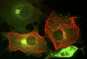
Background imageVesicle Collection: Cell structure, fluorescent micrograph

Cell structure, fluorescent micrograph
Cell structure. Fluorescent light micrograph of cultured cells from a cell line derived from African green monkey kidney cells. Microtubules, part of the cells cytoskeleton, are red

Background imageVesicle Collection: Animal cell processes, artwork

Animal cell processes, artwork
Animal cell processes. Cutaway artwork showing the structures inside an animal cell and four different processes that take place inside it or on its membrane (all marked by magnifying glasses)

Background imageVesicle Collection: Golgi apparatus, artwork

Golgi apparatus, artwork. This structure is an organelle found within eukaryotic cells. It receives proteins and lipids that are synthesised elsewhere on the endoplasmic reticulum

Background imageVesicle Collection: Axon anatomy, diagram

Axon anatomy, diagram
Axon anatomy. Diagram of the anatomical structure of an axon, the main extension (dendrite) of a nerve cell. Cellular components include endoplasmic reticulum (green), neurotubules (white)

Background imageVesicle Collection: Chlamydia, SEM

Chlamydia, SEM
Chlamydia. Coloured scanning electron micrograph (SEM) of chlamydia (Chlamydia sp.) bacteria (green, spherical, centre) in a ruptured cell

Background imageVesicle Collection: Yeast cell, electron tomogram

Yeast cell, electron tomogram image. Yeast cell, Schizosaccharomyces pombe, created using a 3-D electron microscope. This involves firing beams of electrons from many different angles to create


All Professionally Made to Order for Quick Shipping

"Exploring the Intricate World of Vesicles: Unveiling Cellular Secrets" Synapse nerve junction, TEM: Witnessing the intricate communication between neurons at a synapse nerve junction through the lens of a transmission electron microscope (TEM). Rough endoplasmic reticulum, TEM: Peering into the rough endoplasmic reticulum within cells, revealing its role in protein synthesis and transport as captured by a transmission electron microscope (TEM). Skin disorders, artwork: Artistic representation showcasing various skin disorders that involve vesicle formation, highlighting their impact on human health and well-being. Cell types, artwork: A captivating artwork depicting different cell types where vesicles play crucial roles in cellular processes such as secretion and intracellular transport. Golgi apparatus, SEM: Exploring the intricacies of the Golgi apparatus using scanning electron microscopy (SEM), unraveling its vital function in modifying and packaging proteins within vesicles. Nerve synapse, TEM: Delving deep into the mesmerizing world of nerve synapses through high-resolution images obtained from a transmission electron microscope (TEM), shedding light on how vesicles facilitate neurotransmitter release. Fibroblast cell, artwork: An artistic portrayal capturing fibroblast cells involved in wound healing and collagen production while emphasizing their involvement with vesicular transport mechanisms. Development of Tadpole (engraving): A historical engraving illustrating tadpole development stages where they are instrumental in metamorphosis progression from aquatic to terrestrial lifeforms. Bladderwrack - Fucus vesiculosus: Discovering bladderwrack seaweed species like Fucus vesiculosus known for its distinctive air-filled reproductive structures called "vesicles, " which aid buoyancy and reproduction. Eruptive fevers - Colour litho & Plate 6 engraving .