Fraud Blocker Skip to main content

Veterinary Medicine Collection

"Exploring the Intricacies of Veterinary Medicine

Background imageVeterinary Medicine Collection: Deer anatomy, artwork

Deer anatomy, artwork
Deer anatomy. Computer artwork showing the internal anatomy of a deer (family Cervidae). Anatomical features shown here include the skeleton, lungs, intestines, bladder

Background imageVeterinary Medicine Collection: Dog skeleton, artwork F006 / 2267

Dog skeleton, artwork F006 / 2267
Dog skeleton, computer artwork

Background imageVeterinary Medicine Collection: Mouse malaria parasite, SEM

Mouse malaria parasite, SEM
Mouse malaria parasite. Coloured scanning electron micrograph (SEM) of a Plasmodium berghei protozoan (yellow) and red blood cells. P. berghei is the parasite that causes malaria in mice

Background imageVeterinary Medicine Collection: Study of a Fowl Head and Neck, Lateral View, from A Comparative Anatomical Exposition of

Study of a Fowl Head and Neck, Lateral View, from A Comparative Anatomical Exposition of the Structure of the Human
XYC262893 Study of a Fowl Head and Neck, Lateral View, from A Comparative Anatomical Exposition of the Structure of the Human Body with that of a Tiger and a Common Fowl

Background imageVeterinary Medicine Collection: Leopard Body, Lateral View, 1795-1806 (graphite & red chalk)

Leopard Body, Lateral View, 1795-1806 (graphite & red chalk)
XYC261357 Leopard Body, Lateral View, 1795-1806 (graphite & red chalk) by Stubbs, George (1724-1806); 24.8x35.2 cm; Yale Center for British Art, Paul Mellon Collection

Background imageVeterinary Medicine Collection: Study of a Tiger, Lateral View, from A Comparative Anatomical Exposition of the Structure of

Study of a Tiger, Lateral View, from A Comparative Anatomical Exposition of the Structure of the Human Body with that
XYC261343 Study of a Tiger, Lateral View, from A Comparative Anatomical Exposition of the Structure of the Human Body with that of a Tiger and a Common Fowl

Background imageVeterinary Medicine Collection: Study of a Tiger, Standing in Human Posture, from A Comparative Anatomical Exposition of

Study of a Tiger, Standing in Human Posture, from A Comparative Anatomical Exposition of the Structure of the Human
XYC261362 Study of a Tiger, Standing in Human Posture, from A Comparative Anatomical Exposition of the Structure of the Human Body with that of a Tiger and a Common Fowl

Background imageVeterinary Medicine Collection: Study of a Tiger, Recumbent, Viewed Ventrally, from A Comparative Anatomical Exposition of

Study of a Tiger, Recumbent, Viewed Ventrally, from A Comparative Anatomical Exposition of the Structure of the Human
XYC261361 Study of a Tiger, Recumbent, Viewed Ventrally, from A Comparative Anatomical Exposition of the Structure of the Human Body with that of a Tiger and a Common Fowl

Background imageVeterinary Medicine Collection: Study of a Tiger, Lateral View, Diagram Showing the Subcutaneous Blood Supply

Study of a Tiger, Lateral View, Diagram Showing the Subcutaneous Blood Supply
XYC261342 Study of a Tiger, Lateral View, Diagram Showing the Subcutaneous Blood Supply, from A Comparative Anatomical Exposition of the Structure of the Human Body with that of a Tiger

Background imageVeterinary Medicine Collection: Study of a Tiger, Lateral View, from A Comparative Anatomical Exposition of the Structure of

Study of a Tiger, Lateral View, from A Comparative Anatomical Exposition of the Structure of the Human Body with that
XYC261340 Study of a Tiger, Lateral View, from A Comparative Anatomical Exposition of the Structure of the Human Body with that of a Tiger and a Common Fowl

Background imageVeterinary Medicine Collection: Study of a Fowl, Lateral View, from A Comparative Anatomical Exposition of the Structure of

Study of a Fowl, Lateral View, from A Comparative Anatomical Exposition of the Structure of the Human Body with that of
XYC261368 Study of a Fowl, Lateral View, from A Comparative Anatomical Exposition of the Structure of the Human Body with that of a Tiger and a Common Fowl

Background imageVeterinary Medicine Collection: Study of a Fowl, Lateral View, from A Comparative Anatomical Exposition of the Structure of

Study of a Fowl, Lateral View, from A Comparative Anatomical Exposition of the Structure of the Human Body with that of
XYC262888 Study of a Fowl, Lateral View, from A Comparative Anatomical Exposition of the Structure of the Human Body with that of a Tiger and a Common Fowl

Background imageVeterinary Medicine Collection: Study of a Tiger, Lateral View, from A Comparative Anatomical Exposition of the Structure of

Study of a Tiger, Lateral View, from A Comparative Anatomical Exposition of the Structure of the Human Body with that
XYC261339 Study of a Tiger, Lateral View, from A Comparative Anatomical Exposition of the Structure of the Human Body with that of a Tiger and a Common Fowl

Background imageVeterinary Medicine Collection: Study of a Fowl, Lateral View, from A Comparative Anatomical Exposition of the Structure of

Study of a Fowl, Lateral View, from A Comparative Anatomical Exposition of the Structure of the Human Body with that of
XYC261366 Study of a Fowl, Lateral View, from A Comparative Anatomical Exposition of the Structure of the Human Body with that of a Tiger and a Common Fowl

Background imageVeterinary Medicine Collection: Study of a Tiger, Lateral View, from A Comparative Anatomical Exposition of the Structure of

Study of a Tiger, Lateral View, from A Comparative Anatomical Exposition of the Structure of the Human Body with that
XYC261337 Study of a Tiger, Lateral View, from A Comparative Anatomical Exposition of the Structure of the Human Body with that of a Tiger and a Common Fowl

Background imageVeterinary Medicine Collection: Study of a Fowl, Lateral View, from A Comparative Anatomical Exposition of the Structure of

Study of a Fowl, Lateral View, from A Comparative Anatomical Exposition of the Structure of the Human Body with that of
XYC261365 Study of a Fowl, Lateral View, from A Comparative Anatomical Exposition of the Structure of the Human Body with that of a Tiger and a Common Fowl

Background imageVeterinary Medicine Collection: Study of a Tiger, Lateral View, from A Comparative Anatomical Exposition of the Structure of

Study of a Tiger, Lateral View, from A Comparative Anatomical Exposition of the Structure of the Human Body with that
XYC261347 Study of a Tiger, Lateral View, from A Comparative Anatomical Exposition of the Structure of the Human Body with that of a Tiger and a Common Fowl

Background imageVeterinary Medicine Collection: Study of a Tiger, Lateral View, from A Comparative Anatomical Exposition of the Structure of

Study of a Tiger, Lateral View, from A Comparative Anatomical Exposition of the Structure of the Human Body with that
XYC261344 Study of a Tiger, Lateral View, from A Comparative Anatomical Exposition of the Structure of the Human Body with that of a Tiger and a Common Fowl

Background imageVeterinary Medicine Collection: Portrait of William Taplin, author of The Gentlemans Stable Directory

Portrait of William Taplin, author of The Gentlemans Stable Directory, A New Compendium of Farriery, etc
6337429 Portrait of William Taplin, author of The Gentlemans Stable Directory, A New Compendium of Farriery, etc by Scott, J

Background imageVeterinary Medicine Collection: Dorking Hen Body, Lateral View, from A Comparative Anatomical Exposition of

Dorking Hen Body, Lateral View, from A Comparative Anatomical Exposition of
XYC262896 Dorking Hen Body, Lateral View, from A Comparative Anatomical Exposition of the Structure of the Human Body with that of a Tiger and a Common Fowl

Background imageVeterinary Medicine Collection: Dorking Hen Body, Lateral View, from A Comparative Anatomical Exposition of

Dorking Hen Body, Lateral View, from A Comparative Anatomical Exposition of
XYC262895 Dorking Hen Body, Lateral View, from A Comparative Anatomical Exposition of the Structure of the Human Body with that of a Tiger and a Common Fowl, 1795-1806 (pen & ink)

Background imageVeterinary Medicine Collection: Artillery on the Dunes, Flanders, 1915 (w / c)

Artillery on the Dunes, Flanders, 1915 (w / c)
5920312 Artillery on the Dunes, Flanders, 1915 (w/c) by Jones, Adrian (1845-1938); National Army Museum, London; (add.info.: Artillery on the Dunes, Flanders, 1915)

Background imageVeterinary Medicine Collection: The Plague in Animals and Humans (engraving) (b / w photo)

The Plague in Animals and Humans (engraving) (b / w photo)
CHT237284 The Plague in Animals and Humans (engraving) (b/w photo) by Raphael (Raffaello Sanzio of Urbino) (1483-1520) (after); Bibliotheque des Arts Decoratifs, Paris

Background imageVeterinary Medicine Collection: Cow anatomy, artwork

Cow anatomy, artwork
Cow anatomy. Computer artwork showing the internal anatomy of a domesticated cow (Bos primigenius). Anatomical features shown here include the skeleton, lungs, intestines, bladder

Background imageVeterinary Medicine Collection: Dorking Hen Skeleton, Lateral View, from A Comparative Anatomical Exposition

Dorking Hen Skeleton, Lateral View, from A Comparative Anatomical Exposition
XYC262894 Dorking Hen Skeleton, Lateral View, from A Comparative Anatomical Exposition of the Structure of the Human Body with that of a Tiger and a Common Fowl

Background imageVeterinary Medicine Collection: Domestic cat with a neck brace after a visit to the vets

Domestic cat with a neck brace after a visit to the vets

Background imageVeterinary Medicine Collection: Mustang Liniment Trade Card. ca. 1890, Mustang Liniment Trade Card

Mustang Liniment Trade Card. ca. 1890, Mustang Liniment Trade Card

Background imageVeterinary Medicine Collection: Injured dog wearing Elizabethan-style collar round neck

Injured dog wearing Elizabethan-style collar round neck

Background imageVeterinary Medicine Collection: Prising open jaws of cat (Felis silvestris cattus) to examine teeth

Prising open jaws of cat (Felis silvestris cattus) to examine teeth

Background imageVeterinary Medicine Collection: Boxer dog (Canis familiaris) with its leg in plaster

Boxer dog (Canis familiaris) with its leg in plaster

Background imageVeterinary Medicine Collection: A female Mallard (Anas platyrhynchos) with a bandage around its wing

A female Mallard (Anas platyrhynchos) with a bandage around its wing

Background imageVeterinary Medicine Collection: A kitten receiving an injection

A kitten receiving an injection

Background imageVeterinary Medicine Collection: Horse cardiovascular system, artwork F006 / 2351

Horse cardiovascular system, artwork F006 / 2351
Horse cardiovascular system, computer artwork

Background imageVeterinary Medicine Collection: Horse digestive system, artwork

Horse digestive system, artwork
Horse digestive system, computer artwork

Background imageVeterinary Medicine Collection: Dog central nervous system, artwork

Dog central nervous system, artwork
Dog central nervous system, computer artwork

Background imageVeterinary Medicine Collection: Tapeworm head, SEM C014 / 4877

Tapeworm head, SEM C014 / 4877
Tapeworm head. Coloured scanning electron micrograph of the head (scolex) of a tapeworm (class Cestoda) found in a shark, showing the hooks used to attach to the host animals digestive tract

Background imageVeterinary Medicine Collection: Tapeworm head, SEM C014 / 4880

Tapeworm head, SEM C014 / 4880
Tapeworm head. Coloured scanning electron micrograph of the head (scolex) of a tapeworm (Platybothrium auriculatum) found in a blue shark (Prionace glauca)

Background imageVeterinary Medicine Collection: Tapeworm head, SEM C014 / 4878

Tapeworm head, SEM C014 / 4878
Tapeworm head. Coloured scanning electron micrograph of the head (scolex) of a tapeworm (class Cestoda) found in a shark, showing the hooks used to attach to the host animals digestive tract

Background imageVeterinary Medicine Collection: Tapeworm head, SEM C014 / 4879

Tapeworm head, SEM C014 / 4879
Tapeworm head. Coloured scanning electron micrograph of the head (scolex) of a tapeworm (class Cestoda) found in a shark, showing the suckers used to attach to the host animals digestive tract

Background imageVeterinary Medicine Collection: Veterinarians Theiler and Mohler in 1923 C017 / 7824

Veterinarians Theiler and Mohler in 1923 C017 / 7824
Veterinarians Theiler and Mohler in 1923. Swiss-South African veterinarian Arnold Theiler (left, 1867-1936) and US veterinarian John Robbins Mohler (right, 1875-1952)

Background imageVeterinary Medicine Collection: Animal euthanasia, early 20th century C016 / 4578

Animal euthanasia, early 20th century C016 / 4578
Animal euthanasia. Men preparing to euthanase (humanely kill) a dog using an apparatus that produces carbonic acid gas (archaic term for carbon dioxide)

Background imageVeterinary Medicine Collection: Henri Bouley, French veterinarian

Henri Bouley, French veterinarian
Henri Bouley (1814-1885), French veterinarian. Bouley, professor and director at the veterinary school at Maisons-Alfort, was made Inspector-General of veterinary schools in 1866

Background imageVeterinary Medicine Collection: Inoculating chicken eggs

Inoculating chicken eggs. Medical researchers working on treatment for bird viruses. At left, one researcher is marking the eggs, while at right

Background imageVeterinary Medicine Collection: Injured lion, conceptual artwork

Injured lion, conceptual artwork

Background imageVeterinary Medicine Collection: Tapeworm head, SEM

Tapeworm head, SEM
Tapeworm head. Coloured scanning electron micrograph of the head (scolex) of a tapeworm (Taenia pisiformis), showing the hooks used to attach to the host animals digestive tract

Background imageVeterinary Medicine Collection: Dog anatomy, artwork

Dog anatomy, artwork
Dog anatomy. Computer artwork showing the skeletal system of a domesticated dog (Canis lupus familiaris)

Background imageVeterinary Medicine Collection: SARS virus, conceptual artwork

SARS virus, conceptual artwork
SARS virus, conceptual computer artwork. Severe acute respiratory syndrome (SARS) is an often fatal lung disease that first appeared in China in 2002

Background imageVeterinary Medicine Collection: SARS virus and chicken, artwork

SARS virus and chicken, artwork
SARS virus and chicken, computer artwork. Severe acute respiratory syndrome (SARS) is an often fatal lung disease that first appeared in China in 2002


All Professionally Made to Order for Quick Shipping

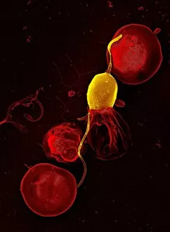
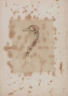
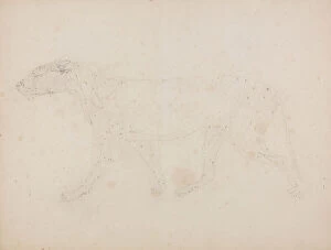
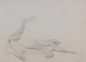
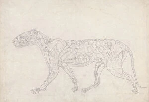
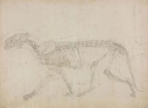
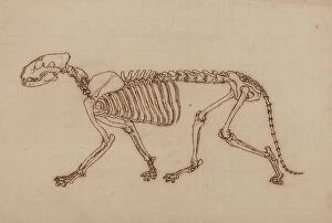
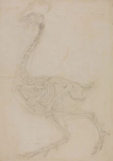
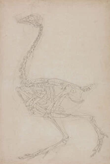
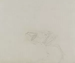
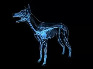
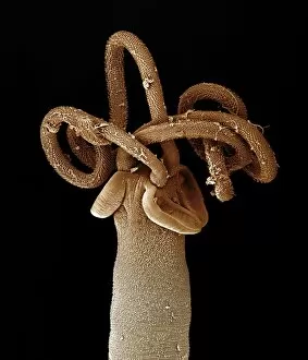
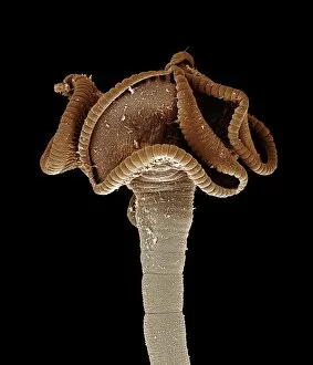
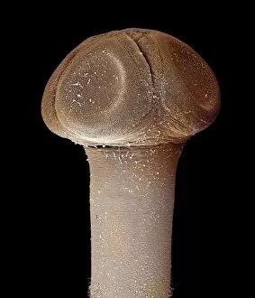
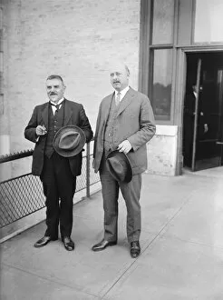
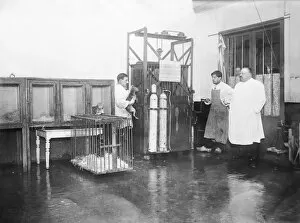
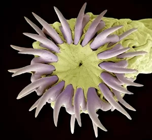
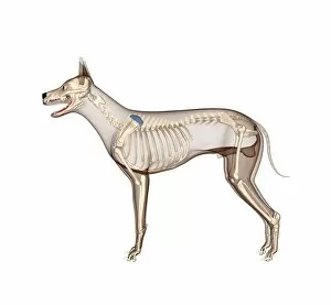
"Exploring the Intricacies of Veterinary Medicine: From Ancient Artworks to Modern Discoveries" Step into the fascinating world as we delve into its rich history and cutting-edge advancements. 🐾 Unearthed from the depths of time, a dog skeleton (artwork F006/2267) serves as a reminder of our enduring bond with animals. It symbolizes the foundation upon which veterinary medicine was built. Zooming in closer, we encounter a microscopic marvel - the mouse malaria parasite captured through a scanning electron microscope (SEM). This image showcases how veterinarians tirelessly combat diseases that affect both animals and humans alike. Moving on to canine anatomy, an intricately detailed artwork sheds light on the inner workings of man's best friend. Such knowledge allows veterinarians to diagnose and treat ailments with precision and care. In another corner, we find ourselves face-to-face with William Taplin, author extraordinaire whose literary contributions have shaped equine health for generations. His work in "The Gentleman's Stable Directory" has become an invaluable resource for horse owners worldwide. Venturing beyond domesticated companions, we explore avian anatomy through captivating illustrations like that of a Dorking Hen's body in lateral view. These comparative anatomical exposés provide insights into diverse species' unique physiological structures. As our journey continues, wartime imagery emerges - artillery on Flanders' dunes during 1915 reminds us that even amidst chaos and conflict, veterinary professionals stood steadfastly by their animal patients' sides. A haunting engraving depicts "The Plague in Animals and Humans, " underscoring veterinarians' crucial role in safeguarding public health against zoonotic diseases throughout history. An antique illustration reveals early experiments conducted by pioneering surgeons who sought to advance veterinary surgery techniques using rabbits as subjects. Their dedication paved the way for modern surgical practices benefiting countless animals today. Delving deeper into nature's wonders, deer and cow anatomy artworks showcase the intricate complexity of these majestic creatures.