Fraud Blocker Skip to main content

Viral Collection (page 8)

"Unveiling the Intricate World of Viruses: From Coronavirus to Norovirus, Hepatitis C to Influenza" In this captivating illustration

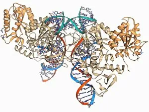
Background imageViral Collection: Retroviral intasome molecule F006 / 9706

Retroviral intasome molecule F006 / 9706
Retroviral intasome molecule. Molecular model of an intasome from a retrovirus complexed with host cell DNA (deoxyribonucleic acid)

Background imageViral Collection: Lassa virus nucleocapsid protein F006 / 9702

Lassa virus nucleocapsid protein F006 / 9702
Lassa virus nucleocapsid protein, molecular model. This protein has essential roles in RNA (ribonucleic acid) synthesis and immune suppression

Background imageViral Collection: Human rotavirus enterotoxin F006 / 9701

Human rotavirus enterotoxin F006 / 9701
Human rotavirus enterotoxin. Molecular model of the NSP4 (nonstructural protein 4) from the human rotavirus. This enterotoxin induces diarrhoea

Background imageViral Collection: Ebola viral protein 35 and RNA F006 / 9697

Ebola viral protein 35 and RNA F006 / 9697
Ebola viral protein 35 and RNA. Molecular model of the Ebola viral protein 35 (VP35) bound to a molecule of double stranded RNA (ribonucleic acid)

Background imageViral Collection: Bluetongue virus capsid, molecular model F006 / 9687

Bluetongue virus capsid, molecular model F006 / 9687
Bluetongue virus capsid, molecular model. This virus, transmitted by midges, causes bluetongue disease in ruminants (commonly sheep, and less frequently cattle, goats and deer)

Background imageViral Collection: Hepatitis E virus capsid, molecular model F006 / 9688

Hepatitis E virus capsid, molecular model F006 / 9688
Hepatitis E virus capsid, molecular model. This virus causes the disease hepatitis E in humans. Transmitted by faecal-oral contact

Background imageViral Collection: HIV reverse transcription enzyme F006 / 9684

HIV reverse transcription enzyme F006 / 9684
HIV reverse transcription enzyme. Molecular model of the reverse transcriptase enzyme found in HIV (the human immunodeficiency virus) bound to the inhibitor nevirapine

Background imageViral Collection: HIV DNA and transcription factor F006 / 9680

HIV DNA and transcription factor F006 / 9680
HIV DNA and transcription factor. Molecular model of DNA (deoxyribonucleic acid) from HIV-1 (human immunodeficiency virus type 1) complexed with the transcription factor kappa B

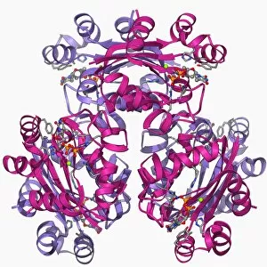
Background imageViral Collection: Nucleoside diphosphate kinase molecule F006 / 9671

Nucleoside diphosphate kinase molecule F006 / 9671
Nucleoside diphosphate kinase. Molecular model of the enzyme nucleoside diphosphate kinase from the Acanthamoeba polyphaga mimivirus

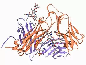
Background imageViral Collection: Interferon antagonism by viral protein F006 / 9662

Interferon antagonism by viral protein F006 / 9662
Interferon (IFN) antagonism by viral protein. Molecular model of an orthopoxvirus IFN-gamma-binding protein bound to an IFN-gamma molecule

Background imageViral Collection: Rous sarcoma virus capsid F006 / 9653

Rous sarcoma virus capsid F006 / 9653
Rous sarcoma virus capsid, molecular model. In viruses, the capsid is the protein shell that encloses the genetic material

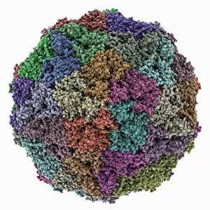
Background imageViral Collection: Grapevine fanleaf virus capsid

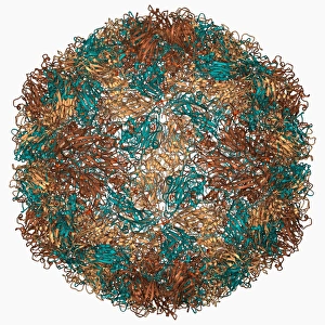
Grapevine fanleaf virus capsid, molecular model. This plant virus is named for its infection of grape vines. It is transmitted by the nematode worm Xiphinema index

Background imageViral Collection: Adenovirus protein and tumor suppressor F006 / 9641

Adenovirus protein and tumor suppressor F006 / 9641
Adenovirus protein and tumour suppressor. Molecular model of the E1A protein from human adenovirus bound to a retinoblastoma tumour suppressor

Background imageViral Collection: Internal ribosome entry site F006 / 9631

Internal ribosome entry site F006 / 9631
Internal ribosome entry site. Molecular model of an internal ribosome entry site nucleotide sequence from the hepatitis C virus. This sequence is essential for the initiation of viral translation

Background imageViral Collection: Mengovirus capsid, molecular model F006 / 9617

Mengovirus capsid, molecular model F006 / 9617
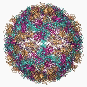
Mengovirus capsid, molecular model. A capsid consists of subunits called capsomeres that self-assemble to form the shell seen here

Background imageViral Collection: HIV antibody therapy, molecular model F006 / 9622

HIV antibody therapy, molecular model F006 / 9622
HIV antibody therapy. Molecular model of the interaction of the HIV surface protein gp120 (green) as it interacts with a human white blood cell surface protein (CD4)

Background imageViral Collection: Viral RNA packaging signal complex F006 / 9609

Viral RNA packaging signal complex F006 / 9609
Viral RNA packaging signal complex. Molecular model of the muPsi RNA packaging signal complex from the Rous sarcoma vuris

Background imageViral Collection: HIV reverse transcription enzyme F006 / 9606

HIV reverse transcription enzyme F006 / 9606
HIV reverse transcription enzyme. Molecular model of the reverse transcriptase enzyme (pink) found in HIV (the human immunodeficiency virus)

Background imageViral Collection: Hepatitis B virus capsid, molecular model F006 / 9594

Hepatitis B virus capsid, molecular model F006 / 9594
Hepatitis B virus capsid, molecular model. This virus, transmitted through infected bodily fluids or blood, causes the disease hepatitis B in humans, leading to acute liver inflammation

Background imageViral Collection: Human rotavirus capsid protein molecule F006 / 9582

Human rotavirus capsid protein molecule F006 / 9582
Human rotavirus capsid protein, molecular model

Background imageViral Collection: Adenovirus penton base protein F006 / 9572

Adenovirus penton base protein F006 / 9572
Adenovirus penton base protein, molecular model. This protein molecule is a subunit called a penton, forming the vertices of the capsid of this adenovirus

Background imageViral Collection: Flu virus surface protein molecule F006 / 9566

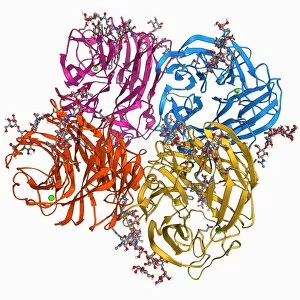
Flu virus surface protein molecule F006 / 9566
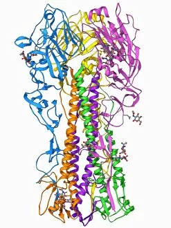
Flu virus surface protein molecule. Molecular model of the neuraminidase glycoprotein enzyme found on the surface of the influenza A (flu) virus

Background imageViral Collection: SARS virus surface protein molecule F006 / 9560

SARS virus surface protein molecule F006 / 9560
SARS virus surface protein. Molecular model of the spike 2 (S2) envelope glycoprotein from the severe acute respiratory syndrome (SARS) virus

Background imageViral Collection: RNA stem-loop motif, molecular model F006 / 9544

RNA stem-loop motif, molecular model F006 / 9544
RNA stem-loop motif. Molecular model of the stem-loop II motif from the SARS (severe acute respiratory syndrome) coronavirus. This RNA (ribonucleic acid) element is a target for antiviral drugs

Background imageViral Collection: SV40 virus capsid, molecular model F006 / 9508

SV40 virus capsid, molecular model F006 / 9508
SV40 virus capsid, molecular model. Simian virus 40 (SV40) is found in monkeys such as Rhesus monkeys and macaques. Potentially tumour-causing, it is used in laboratory research and in vaccines

Background imageViral Collection: Reverse transcriptase and inhibitor F006 / 9519

Reverse transcriptase and inhibitor F006 / 9519
Reverse transcriptase and inhibitor. Molecular model of HIV reverse transcriptase complexed with a non-nucleoside reverse transcriptase inhibitor drug

Background imageViral Collection: T cell receptor, molecular model F006 / 9515

T cell receptor, molecular model F006 / 9515
T cell receptor. Molecular model of an alpha T cell receptor. T cell receptors are protein complexes found on the surface of a type of white blood cell called T lymphocytes (or T cells)

Background imageViral Collection: DNA helicase molecule F006 / 9509

DNA helicase molecule F006 / 9509
DNA helicase. Molecular model of a helicase molecule from the SV40 virus. Helicases are enzymes that separate the two strands of the DNA double helix

Background imageViral Collection: Simian virus SV40 large T antigen F006 / 9513

Simian virus SV40 large T antigen F006 / 9513
Simian virus (SV40) large T antigen, molecular model. This antigen is from the simian vacuolating virus 40 (SV40). Large T antigens play a role in regulating the viral life cycle of

Background imageViral Collection: Parvovirus particle, molecular model F006 / 9499

Parvovirus particle, molecular model F006 / 9499
Parvovirus particle. Molecular model showing the structure of the capsid (outer protein coat) of a human parvovirus (family Parvoviridae) particle

Background imageViral Collection: SARS virus capsid protein F006 / 9504

SARS virus capsid protein F006 / 9504
SARS virus capsid protein, molecular model. This protein is responsible for binding the capsid (outer coat) of the SARS (severe acute respiratory syndrome)

Background imageViral Collection: Rhinovirus capsid, molecular model F006 / 9490

Rhinovirus capsid, molecular model F006 / 9490
Rhinovirus capsid, molecular model. This is human rhinovirus. The rhinovirus infects the upper respiratory tract and is the cause of the common cold. It is spread by coughs and sneezes

Background imageViral Collection: H1 antigen from 1918 influenza virus F006 / 9495

H1 antigen from 1918 influenza virus F006 / 9495
H1 antigen from 1918 influenza virus

Background imageViral Collection: RNA interference viral suppressor and RNA F006 / 9488

RNA interference viral suppressor and RNA F006 / 9488
RNA interference viral suppressor and RNA. Molecular model of the p19 protein (yellow) from a Tombusvirus, suppressing a double-stranded, small interfering RNA (siRNA) molecule (red and blue)

Background imageViral Collection: Foot-and-mouth disease virus F006 / 9471

Foot-and-mouth disease virus F006 / 9471
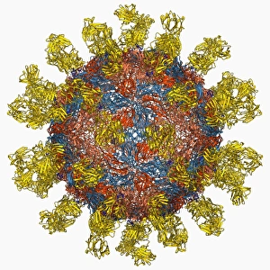
Foot-and-mouth disease virus. Molecular model of the foot-and-mouth disease (FMD) virus Aphtae epizooticae, with antibodies (immunoglobulins)

Background imageViral Collection: Herpesvirus immune inhibitor protein F006 / 9476

Herpesvirus immune inhibitor protein F006 / 9476
Herpesvirus immune inhibitor protein. Molecular model of the protein ICP47 from the herpes simplex virus-1 (HSV-1). This protein prevents antigens from the virus being presented to immune cells

Background imageViral Collection: Poliovirus type 3 capsid, molecular model F006 / 9465

Poliovirus type 3 capsid, molecular model F006 / 9465
Poliovirus type 3 capsid, molecular model. This enterovirus causes poliomyelitis (polio) in humans, which affects the nervous system, sometimes leading to paralysis

Background imageViral Collection: Adenovirus fibre shaft protein F006 / 9473

Adenovirus fibre shaft protein F006 / 9473
Adenovirus fibre shaft protein, molecular model. This protein forms part of the shaft of the fibres or spikes by which adenoviruses attach to receptors on cells during the infection process

Background imageViral Collection: Adenovirus hexon protein F006 / 9453

Adenovirus hexon protein F006 / 9453
Adenovirus hexon protein, molecular model. Hexon proteins are part of the protein coat or shell (capsid) of adenoviruses. In viruses

Background imageViral Collection: Dengue virus surface protein molecule F006 / 9449

Dengue virus surface protein molecule F006 / 9449
Dengue virus surface protein molecule. Molecular model of the envelope glycoprotein found on the surface of the dengue haemorrhagic fever (DHF) virus

Background imageViral Collection: Human poliovirus, molecular model F006 / 9434

Human poliovirus, molecular model F006 / 9434
Human poliovirus particle. Molecular model of the capsid of the human poliovirus. The capsid is a protein coat that encloses the viruss genetic information (genome), stored as RNA (ribonucleic acid)

Background imageViral Collection: VEE equine encephalitis virus capsid

VEE equine encephalitis virus capsid
Venezuelan equine encephalitis virus capsid, molecular model. This mosquito-borne virus can kill horses and other equine species, causing brain and spinal cord inflammation

Background imageViral Collection: Herpesvirus capsid protein F006 / 9435

Herpesvirus capsid protein F006 / 9435
Herpesvirus capsid protein. Molecular model of the major capsid protein VP5 from herpes simplex virus-1 (HSV-1). This virus infects epithelial cells around the mouth

Background imageViral Collection: Rhinovirus 14 capsid, molecular model F006 / 9430

Rhinovirus 14 capsid, molecular model F006 / 9430
Rhinovirus 14 capsid, molecular model. This is human rhinovirus 14. The rhinovirus infects the upper respiratory tract and is the cause of the common cold. It is spread by coughs and sneezes

Background imageViral Collection: Flu virus surface protein molecule F006 / 9433

Flu virus surface protein molecule F006 / 9433
Flu virus surface protein molecule. Molecular model of the neuraminidase glycoprotein enzyme found on the surface of the influenza A (flu) virus

Background imageViral Collection: Flu virus surface protein and antibody F006 / 9428

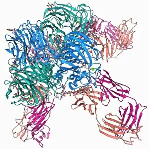
Flu virus surface protein and antibody F006 / 9428
Flu virus surface protein and antibody. Molecular model of the neuraminidase glycoprotein enzyme found on the surface of the influenza (flu) virus

Background imageViral Collection: DNA helicase molecule F006 / 9426

DNA helicase molecule F006 / 9426
DNA helicase. Molecular model of a helicase molecule from the SV40 virus. Helicases are enzymes that separate the two strands of the DNA double helix

Background imageViral Collection: Hepatitis C polymerase enzyme F006 / 9427

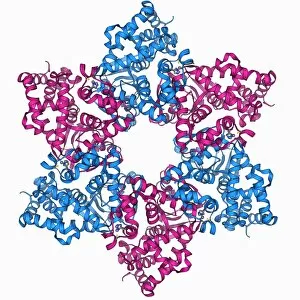
Hepatitis C polymerase enzyme F006 / 9427
Hepatitis C polymerase enzyme, molecular model. This protein is the NS5b RNA polymerase found in the virus hepatitis C (genotype-1b, strain J4)


All Professionally Made to Order for Quick Shipping

"Unveiling the Intricate World of Viruses: From Coronavirus to Norovirus, Hepatitis C to Influenza" In this captivating illustration, we delve into the microscopic realm of viruses that have left an indelible mark on our lives. Firstly, we encounter a mesmerizing depiction of the Coronavirus Structure Illustration. Its intricate spikes and spherical shape remind us of its global impact and ongoing battle against humanity. Moving forward, we witness Norovirus particles through a powerful TEM image. These tiny entities are notorious for causing stomach upsets and outbreaks in various settings. Next, our attention is drawn towards a molecular model showcasing the Hepatitis C virus enzyme. This visual representation highlights the complexity behind this viral infection that affects millions worldwide. As we explore further, an artistic rendering presents Flu virus particle artwork F008/3245. It serves as a reminder of how influenza can swiftly spread across populations during seasonal outbreaks. The dangers posed by infections transmitted through sneezing are vividly depicted in artwork C013/5949. The visualization captures the moment when pathogens disperse into the air with every forceful expulsion from an infected individual's respiratory system. Returning to coronavirus territory, another TEM image showcases Coronavirus particles in all their glory. Their distinctive crown-like appearance reinforces their notoriety as agents responsible for severe respiratory illnesses like SARS and MERS. Shifting gears slightly, an artistic portrayal introduces us to Adenovirus - known for causing respiratory infections but also harnessed as vectors in gene therapy research due to their unique properties. Our journey continues with yet another TEM image revealing Influenza virus particles - these minuscule culprits have been responsible for numerous pandemics throughout history and continue to pose significant health threats today. Stepping away momentarily from respiratory viruses, we observe Herpes virus replicating within cells under computer-generated artwork. This glimpse into its life cycle reminds us of the persistence and challenges associated with managing this widespread infection.