Fraud Blocker Skip to main content

Virion Collection (page 8)

A virion is a fascinating microscopic entity that plays a crucial role in the world of viruses and infectious diseases

Background imageVirion Collection: SV40 virus capsid, molecular model F006 / 9508

SV40 virus capsid, molecular model F006 / 9508
SV40 virus capsid, molecular model. Simian virus 40 (SV40) is found in monkeys such as Rhesus monkeys and macaques. Potentially tumour-causing, it is used in laboratory research and in vaccines

Background imageVirion Collection: Parvovirus particle, molecular model F006 / 9499

Parvovirus particle, molecular model F006 / 9499
Parvovirus particle. Molecular model showing the structure of the capsid (outer protein coat) of a human parvovirus (family Parvoviridae) particle

Background imageVirion Collection: Rhinovirus capsid, molecular model F006 / 9490

Rhinovirus capsid, molecular model F006 / 9490
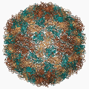
Rhinovirus capsid, molecular model. This is human rhinovirus. The rhinovirus infects the upper respiratory tract and is the cause of the common cold. It is spread by coughs and sneezes

Background imageVirion Collection: Foot-and-mouth disease virus F006 / 9471

Foot-and-mouth disease virus F006 / 9471
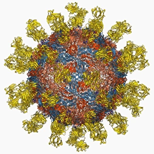
Foot-and-mouth disease virus. Molecular model of the foot-and-mouth disease (FMD) virus Aphtae epizooticae, with antibodies (immunoglobulins)

Background imageVirion Collection: Poliovirus type 3 capsid, molecular model F006 / 9465

Poliovirus type 3 capsid, molecular model F006 / 9465
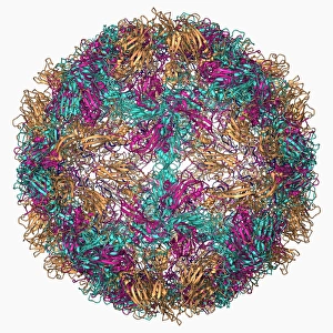
Poliovirus type 3 capsid, molecular model. This enterovirus causes poliomyelitis (polio) in humans, which affects the nervous system, sometimes leading to paralysis

Background imageVirion Collection: Dengue virus capsid, molecular model F006 / 9455

Dengue virus capsid, molecular model F006 / 9455
Dengue virus capsid, molecular model. This virus, transmitted by mosquito bites, causes the tropical disease dengue fever in humans

Background imageVirion Collection: Rous sarcoma virus capsid protein F006 / 9456

Rous sarcoma virus capsid protein F006 / 9456
Rous sarcoma virus capsid protein. Molecular model of the N-terminal domain of the Rous sarcoma virus capsid protein

Background imageVirion Collection: Nodamura virus capsid, molecular model F006 / 9438

Nodamura virus capsid, molecular model F006 / 9438
Nodamura virus capsid, molecular model. This virus can cause lethal infections in both mammals and insects. In viruses, the capsid is the protein shell that encloses the genetic material

Background imageVirion Collection: Human poliovirus, molecular model F006 / 9434

Human poliovirus, molecular model F006 / 9434
Human poliovirus particle. Molecular model of the capsid of the human poliovirus. The capsid is a protein coat that encloses the viruss genetic information (genome), stored as RNA (ribonucleic acid)

Background imageVirion Collection: VEE equine encephalitis virus capsid

VEE equine encephalitis virus capsid
Venezuelan equine encephalitis virus capsid, molecular model. This mosquito-borne virus can kill horses and other equine species, causing brain and spinal cord inflammation

Background imageVirion Collection: Rhinovirus 14 capsid, molecular model F006 / 9430

Rhinovirus 14 capsid, molecular model F006 / 9430
Rhinovirus 14 capsid, molecular model. This is human rhinovirus 14. The rhinovirus infects the upper respiratory tract and is the cause of the common cold. It is spread by coughs and sneezes

Background imageVirion Collection: Bacteriophage alpha 3 capsid F006 / 9414

Bacteriophage alpha 3 capsid F006 / 9414
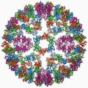
Bacteriophage alpha 3 capsid, molecular model. Bacteriophages are viruses that infect bacteria. Bacteriophage alpha 3 has a capsid consisting of 60 coat proteins (blue) and 12 pentameric (5-subunit)

Background imageVirion Collection: Dengue virus capsid, molecular model F006 / 9392

Dengue virus capsid, molecular model F006 / 9392
Dengue virus capsid, molecular model. This virus, transmitted by mosquito bites, causes the tropical disease dengue fever in humans

Background imageVirion Collection: Coxsackie B3 virus particle F006 / 9381

Coxsackie B3 virus particle F006 / 9381
Coxsackie B3 virus. Computer model of the capsid of the Coxsackie B3 virus

Background imageVirion Collection: Norwalk virus capsid, molecular model F006 / 9372

Norwalk virus capsid, molecular model F006 / 9372
Norwalk virus capsid, molecular model. This norovirus, which causes a viral form of gastroenteritis, is transmitted from person-to-person or through contaminated food

Background imageVirion Collection: Poliovirus particle F006 / 9306

Poliovirus particle F006 / 9306
Poliovirus particle, molecular model

Background imageVirion Collection: Semliki forest virus capsid F006 / 9297

Semliki forest virus capsid F006 / 9297
Semliki forest virus capsid, molecular model. This virus, named for the forest in Uganda where it was identified, is spread by the bite of mosquitoes. It can infect both humans and animals

Background imageVirion Collection: Human poliovirus, molecular model F006 / 9289

Human poliovirus, molecular model F006 / 9289
Human poliovirus particle. Molecular model of the capsid of the human poliovirus. The capsid is a protein coat that encloses the viruss genetic information (genome), stored as RNA (ribonucleic acid)

Background imageVirion Collection: H1N1 flu virus particle, artwork F006 / 9186

H1N1 flu virus particle, artwork F006 / 9186
H1N1 flu virus particle. Computer artwork of an H1N1 influenza A (flu) virus particle (virion). In the particles lipid envelope (purple) are two types of protein spike

Background imageVirion Collection: Murine minute virus capsid

Murine minute virus capsid, molecular model. This parvovirus infects mice, its only known natural host. It is highly infectious, transmitted through the nose and mouth

Background imageVirion Collection: SV40 virus capsid, molecular model C018 / 7904

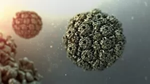
SV40 virus capsid, molecular model C018 / 7904
SV40 virus capsid, molecular model. Simian virus 40 (SV40) is found in monkeys such as Rhesus monkeys and macaques. Potentially tumour-causing, it is used in laboratory research and in vaccines

Background imageVirion Collection: SV40 virus capsid, molecular model C018 / 7903

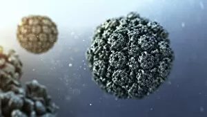
SV40 virus capsid, molecular model C018 / 7903
SV40 virus capsid, molecular model. Simian virus 40 (SV40) is found in monkeys such as Rhesus monkeys and macaques. Potentially tumour-causing, it is used in laboratory research and in vaccines

Background imageVirion Collection: Antibodies and viruses, artwork

Antibodies and viruses, artwork
Antibodies and viruses. Computer artwork showing antibody, or immunoglobulin, molecules (blue) surrounding virus particles (virions, yellow)

Background imageVirion Collection: Reovirus particles, TEM C016 / 9443

Reovirus particles, TEM C016 / 9443
Reovirus particles. Coloured transmission electron micrograph (TEM) of numerous reovirus particles (virions, round). Reoviruses affect the gastrointestinal system and respiratory tract

Background imageVirion Collection: Bacteriophage P22, artwork C016 / 9334

Bacteriophage P22, artwork C016 / 9334
Bacteriophage P22. Computer artwork of an enterobacteria phage P22 particle. A bacteriophage, or phage, is a virus that infects bacteria. Enterobacteria P22 infects Salmonella typhimurium bacteria

Background imageVirion Collection: Rabies virus particle, artwork C016 / 9158

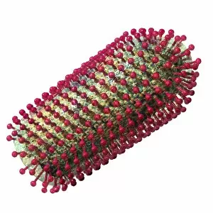
Rabies virus particle, artwork C016 / 9158
Rabies virus particle, computer artwork. This bullet-shaped particle consists of an RNA (ribonucleic acid) genome enclosed in protein capsid

Background imageVirion Collection: Measles virus particle, artwork C016 / 9329

Measles virus particle, artwork C016 / 9329
Measles virus particle, computer artwork. This virus, from the Morbillivirus group of viruses, consists of an RNA (ribonucleic acid)

Background imageVirion Collection: Virus, artwork C016 / 9004

Virus, artwork C016 / 9004
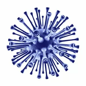
Computer artwork of a generic virus particle, depicting virus types like SARS, MERS and corona

Background imageVirion Collection: Virus, artwork C016 / 9010

Virus, artwork C016 / 9010
Computer artwork of a generic virus particle, depicting virus types like SARS, MERS and corona

Background imageVirion Collection: Bacteriophage T4, artwork C016 / 8968

Bacteriophage T4, artwork C016 / 8968
Bacteriophage T4. Computer artwork of an enterobacteria phage T4 particle. A bacteriophage, or phage, is a virus that infects bacteria. Enterobacteria T4 infects Escherichia coli bacteria

Background imageVirion Collection: Yellow fever virus particles, TEM C016 / 9468

Yellow fever virus particles, TEM C016 / 9468
Yellow fever virus particles. Transmission electron micrograph (TEM) of yellow fever virus particles (virions, yellow). Yellow fever is endemic in tropical Africa and South America

Background imageVirion Collection: Rabies virus particle, artwork C016 / 9157

Rabies virus particle, artwork C016 / 9157
Rabies virus particle, computer artwork. This bullet-shaped particle consists of an RNA (ribonucleic acid) genome enclosed in protein capsid

Background imageVirion Collection: Hepatitis B virus particle, artwork C016 / 9129

Hepatitis B virus particle, artwork C016 / 9129
Hepatitis B virus (HBV) particle, computer artwork. Each particle consists of an outer lipid envelope (green) that surrounds a protein nucleopcapsid (purple)

Background imageVirion Collection: Lassa virus particles, TEM C016 / 9409

Lassa virus particles, TEM C016 / 9409
Lassa virus particles. Transmission electron micrograph (TEM) of Lassa virus particles (virions, blue) amongst cell debris. This Arenavirus is the cause of Lassa fever

Background imageVirion Collection: HIV particles, artwork C016 / 9141

HIV particles, artwork C016 / 9141
HIV particles, computer artwork. HIV (human immunodeficiency virus) causes AIDS (acquired immune deficiency syndrome). The virus consists of an RNA (ribonucleic acid) genome (pink)

Background imageVirion Collection: Flu virus, conceptual artwork C016 / 7530

Flu virus, conceptual artwork C016 / 7530
Flu virus, conceptual computer artwork

Background imageVirion Collection: Adenovirus, artwork C016 / 8960

Adenovirus, artwork C016 / 8960
Adenovirus. Computer artwork showing the external protein structure of an adenovirus particle. Adenoviruses are the largest viruses not to have a protein coat covering their capsid

Background imageVirion Collection: Hepatitis B virus particle, artwork C016 / 9126

Hepatitis B virus particle, artwork C016 / 9126
Hepatitis B virus (HBV) particle, computer artwork. Each particle consists of an outer lipid envelope (green) that surrounds a protein nucleopcapsid (purple)

Background imageVirion Collection: Yellow fever virus particles, TEM C016 / 9469

Yellow fever virus particles, TEM C016 / 9469
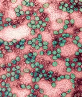
Yellow fever virus particles. Transmission electron micrograph (TEM) of yellow fever virus particles (virions, green). Yellow fever is endemic in tropical Africa and South America

Background imageVirion Collection: Adenovirus, artwork C016 / 8961

Adenovirus, artwork C016 / 8961
Adenovirus. Computer artwork showing the external protein structure of an adenovirus particle. Adenoviruses are the largest viruses not to have a protein coat covering their capsid

Background imageVirion Collection: Adenovirus particles, artwork C016 / 8963

Adenovirus particles, artwork C016 / 8963
Adenovirus particles. Computer artwork showing the external protein structure of adenovirus particles. Adenoviruses are the largest viruses not to have a protein coat covering their capsid

Background imageVirion Collection: Human adenovirus 36, artwork C016 / 8966

Human adenovirus 36, artwork C016 / 8966
Human adenovirus 36. Computer artwork of human adenovirus 36 (HAdV-36), showing the surface structure of the viruss outer protein coat (capsid)

Background imageVirion Collection: Adenovirus particles, artwork C016 / 8965

Adenovirus particles, artwork C016 / 8965
Adenovirus particles. Computer artwork showing the external protein structure of adenovirus particles. Adenoviruses are the largest viruses not to have a protein coat covering their capsid

Background imageVirion Collection: SARS virus particles, TEM C016 / 9445

SARS virus particles, TEM C016 / 9445
SARS virus particles. Coloured transmission electron micrograph (TEM) of a section through a tissue sample infected with numerous SARS coronavirus particles (virions, dark, round)

Background imageVirion Collection: Polio virus particles, TEM C016 / 9436

Polio virus particles, TEM C016 / 9436
Poliovirus particles. Coloured transmission electron micrograph (TEM) of poliovirus particles (round). Poliovirus infects children causing the disease poliomyelitis, or infantile paralysis

Background imageVirion Collection: Acute haemorrhagic conjunctivitis, TEM C016 / 9387

Acute haemorrhagic conjunctivitis, TEM C016 / 9387
Acute haemorrhagic conjunctivitis. Transmission electron micrograph (TEM) of a sample of tissue from the conjunctiva of a human eye that is infected by enterovirus particles (purple)

Background imageVirion Collection: Bacteriophage P22, artwork C016 / 9333

Bacteriophage P22, artwork C016 / 9333
Bacteriophage P22. Computer artwork of an enterobacteria phage P22 particle. A bacteriophage, or phage, is a virus that infects bacteria. Enterobacteria P22 infects Salmonella typhimurium bacteria

Background imageVirion Collection: Rift Valley fever virus particle, artwork C016 / 9160

Rift Valley fever virus particle, artwork C016 / 9160
Rift Valley fever virus particle, computer artwork. This virus particle consists of an RNA (ribonucleic acid) genome enclosed in a protein capsid (yellow)


All Professionally Made to Order for Quick Shipping

A virion is a fascinating microscopic entity that plays a crucial role in the world of viruses and infectious diseases. From the avian flu virus to HIV particles, norovirus particles, and hepatitis B viruses, these tiny structures are responsible for causing widespread infections. Under the powerful lens of a transmission electron microscope (TEM), we can observe the intricate details of various virions. The avian flu virus appears as an ominous presence with its spiky outer surface, while HIV particles reveal their complex structure that enables them to invade human immune cells. Norovirus particles captured by TEM showcase their round shape and distinctive pattern, reminding us of the havoc they wreak on our digestive system during outbreaks. Sneezing becomes more alarming when we realize it can spread infections like wildfire, as depicted in artwork C013 / 5949. Coronavirus particles also make an appearance under TEM; their crown-like spikes serve as a reminder of how this family of viruses has brought about global health crises such as SARS-CoV-2. Adenoviruses stand out with their geometric shapes portrayed in stunning artwork. Hepatitis B viruses take center stage once again due to their persistence and ability to cause chronic liver disease. Computer-generated artwork showcases influenza virus' unique structure while TEM images capture its spherical form along with other strains. Studying virions provides valuable insights into understanding infectious diseases and developing effective countermeasures against them. These captivating entities remind us of the constant battle between humans and pathogens at a microscopic level.