
Virological Collection
"Unveiling the Intricate World of Virology: Exploring Coronavirus, Cold Sores, and More" In this captivating journey into the realm of virology


All Professionally Made to Order for Quick Shipping
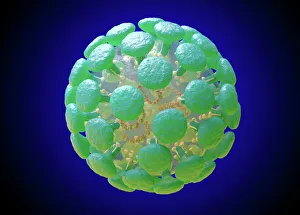
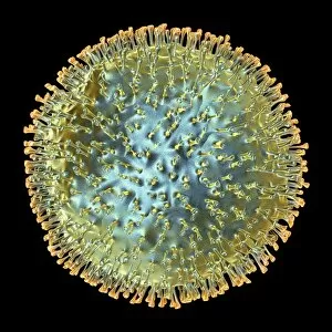
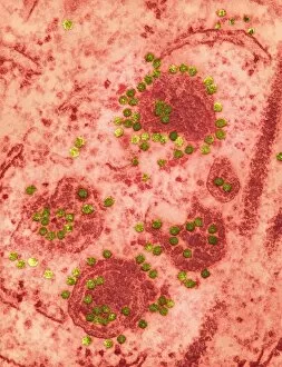
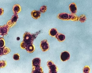
"Unveiling the Intricate World of Virology: Exploring Coronavirus, Cold Sores, and More" In this captivating journey into the realm of virology, we delve deep into the microscopic world of viruses. The first stop on our adventure is a stunning illustration showcasing the intricate structure of the notorious Coronavirus. Its spiky exterior serves as a visual reminder of its ability to infiltrate and wreak havoc within our bodies. Moving forward, we encounter an image depicting a cold sore caused by C014 / 4604 virus. This common yet pesky infection reminds us that even seemingly harmless viruses can disrupt our daily lives. Continuing our exploration, we come across another remarkable illustration revealing the complex architecture of Parvovirus particles (artwork C013 / 4640). These tiny entities possess immense power to invade host cells and replicate themselves with astonishing efficiency. Next up is a conceptual image illustrating flu virus infection. It vividly captures how these elusive pathogens latch onto human cells, causing widespread illness and discomfort during flu seasons. Our journey takes an intriguing turn as we encounter molecular models showcasing HIV enzyme proteins (C014 / 0876). These structures shed light on the mechanisms behind HIV's ability to evade our immune system and persistently infect individuals worldwide. As we near the end of our expedition, avian influenza viruses take center stage in breathtaking artworks (F007 / 0954 - F007 / 0949). These vibrant illustrations remind us of nature's delicate balance and serve as cautionary reminders about potential pandemics that may arise from animal reservoirs. In this mesmerizing voyage through virological wonders, we gain insight into both familiar viral foes like cold sores and influenza while also delving deeper into lesser-known threats such as coronavirus and avian influenza. As scientists continue their tireless efforts to understand these minuscule adversaries better, let us appreciate their complexity while remaining vigilant against their potential harm.