Fraud Blocker Skip to main content

Virology Collection (page 9)

Virology, the fascinating study of viruses and their impact on living organisms

Background imageVirology Collection: Virus particles, artwork F007 / 8504

Virus particles, artwork F007 / 8504
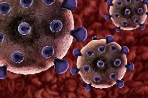
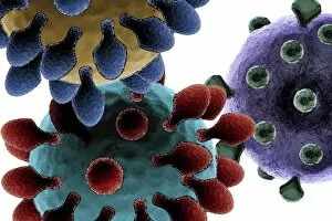
Virus particles, computer artwork

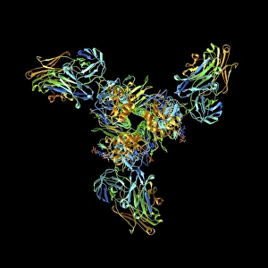
Background imageVirology Collection: Haemagglutinin viral surface protein F007 / 9932

Haemagglutinin viral surface protein F007 / 9932
Haemagglutinin viral surface protein. Molecular model of haemagglutinin, a surface protein from the influenza virus, complexed with a neutralising antibody

Background imageVirology Collection: Virus particles, artwork F007 / 8501

Virus particles, artwork F007 / 8501
Virus particles, computer artwork

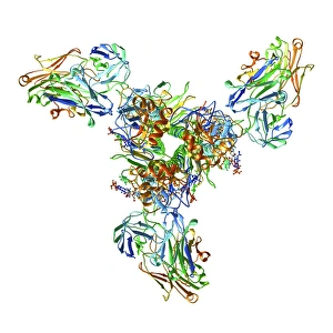
Background imageVirology Collection: Haemagglutinin viral surface protein F007 / 9931

Haemagglutinin viral surface protein F007 / 9931
Haemagglutinin viral surface protein. Molecular model of haemagglutinin, a surface protein from the influenza virus, complexed with a neutralising antibody

Background imageVirology Collection: Virus particles, artwork F007 / 8509

Virus particles, artwork F007 / 8509
Virus particles, computer artwork

Background imageVirology Collection: Virus particles, artwork F007 / 8498

Virus particles, artwork F007 / 8498
Virus particles, computer artwork

Background imageVirology Collection: Virus particles, artwork F007 / 8508

Virus particles, artwork F007 / 8508
Virus particles, computer artwork

Background imageVirology Collection: Viral research, conceptual image F007 / 8519

Viral research, conceptual image F007 / 8519
Viral research, conceptual composite image

Background imageVirology Collection: Viral vaccine, conceptual artwork F007 / 8511

Viral vaccine, conceptual artwork F007 / 8511
Viral vaccine, conceptual computer artwork

Background imageVirology Collection: TEM of a cluster of corona viruses F007 / 9886

TEM of a cluster of corona viruses F007 / 9886
Corona viruses. Coloured transmission electron micrograph of a section through a cluster of corona viruses. The corona viruses cause the common cold, gastroenteritis

Background imageVirology Collection: Viral vaccine, conceptual artwork F007 / 8502

Viral vaccine, conceptual artwork F007 / 8502
Viral vaccine, conceptual computer artwork

Background imageVirology Collection: Virus, artwork F007 / 8500

Virus, artwork F007 / 8500
Virus, computer artwork

Background imageVirology Collection: Virus particles, artwork F007 / 8510

Virus particles, artwork F007 / 8510
Virus particles, computer artwork

Background imageVirology Collection: Virus particles, artwork F007 / 8497

Virus particles, artwork F007 / 8497
Virus particles, computer artwork

Background imageVirology Collection: Virus particles, artwork F007 / 8507

Virus particles, artwork F007 / 8507
Virus particles, computer artwork

Background imageVirology Collection: Virus particles, artwork F007 / 8499

Virus particles, artwork F007 / 8499
Virus particles, computer artwork

Background imageVirology Collection: TEM of a cluster of corona viruses F007 / 9890

TEM of a cluster of corona viruses F007 / 9890
Corona viruses. Coloured transmission electron micrograph of a section through a cluster of corona viruses. The corona viruses cause the common cold, gastroenteritis

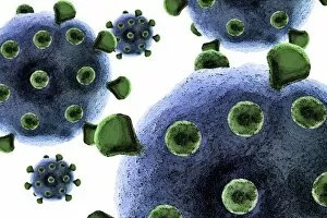
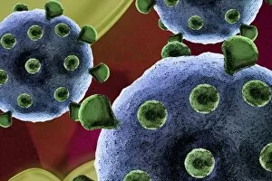
Background imageVirology Collection: Flu virus particles, artwork F008 / 3244

Flu virus particles, artwork F008 / 3244
Flu virus particles, computer artwork. In the virus envelope are two types of protein spike, haemagglutinin (H, light green) and neuraminidase (N, dark green), which determine the strain of virus

Background imageVirology Collection: Virus particles, artwork F008 / 3378

Virus particles, artwork F008 / 3378
Virus particles, computer artwork

Background imageVirology Collection: Flu virus particles, artwork F008 / 3255

Flu virus particles, artwork F008 / 3255
Flu virus particles, computer artwork

Background imageVirology Collection: Virus particles, artwork F008 / 3377

Virus particles, artwork F008 / 3377
Virus particles, computer artwork

Background imageVirology Collection: Flu virus particles, artwork F008 / 3251

Flu virus particles, artwork F008 / 3251
Flu virus particles, computer artwork

Background imageVirology Collection: Virus, artwork F008 / 3374

Virus, artwork F008 / 3374
Virus, computer artwork

Background imageVirology Collection: Flu virus particles, artwork F008 / 3242

Flu virus particles, artwork F008 / 3242
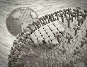
Flu virus particles, computer artwork. In the virus envelope are two types of protein spike, haemagglutinin (H, peg-like) and neuraminidase (N, squares), which determine the strain of virus

Background imageVirology Collection: Flu infection, artwork F008 / 3256

Flu infection, artwork F008 / 3256
Flu infection, computer artwork. Influenza virus particles entering the lungs

Background imageVirology Collection: Flu virus particles, artwork F008 / 3248

Flu virus particles, artwork F008 / 3248
Flu virus particles, computer artwork

Background imageVirology Collection: Flu virus particles, artwork F008 / 3250

Flu virus particles, artwork F008 / 3250
Flu virus particles, computer artwork

Background imageVirology Collection: Flu virus particles, artwork F008 / 3247

Flu virus particles, artwork F008 / 3247
Flu virus particles, computer artwork

Background imageVirology Collection: Flu virus particles, artwork F008 / 3243

Flu virus particles, artwork F008 / 3243
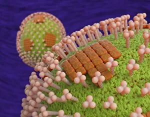
Flu virus particles, computer artwork. In the virus envelope are two types of protein spike, haemagglutinin (H, pink) and neuraminidase (N, orange), which determine the strain of virus

Background imageVirology Collection: Flu virus particles, artwork F008 / 3249

Flu virus particles, artwork F008 / 3249
Flu virus particles, computer artwork

Background imageVirology Collection: Flu virus particles, artwork F008 / 3252

Flu virus particles, artwork F008 / 3252
Flu virus particles, computer artwork

Background imageVirology Collection: Flu virus particles, artwork F008 / 3253

Flu virus particles, artwork F008 / 3253
Flu virus particles, computer artwork

Background imageVirology Collection: Virus particles, artwork F008 / 3376

Virus particles, artwork F008 / 3376
Virus particles, computer artwork

Background imageVirology Collection: Flu virus particles, artwork F008 / 3241

Flu virus particles, artwork F008 / 3241
Flu virus particles, computer artwork. In the virus envelope are two types of protein spike, haemagglutinin (H, peg-like) and neuraminidase (N, squares), which determine the strain of virus

Background imageVirology Collection: Flu virus particles, artwork F008 / 3254

Flu virus particles, artwork F008 / 3254
Flu virus particles, computer artwork

Background imageVirology Collection: Virus particles, artwork F008 / 3375

Virus particles, artwork F008 / 3375
Virus particles, computer artwork

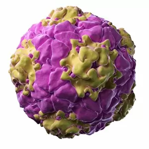
Background imageVirology Collection: L-A virus, artwork

L-A virus, artwork
L-A virus, computer artwork. This virus infects yeast cells

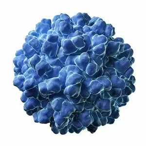
Background imageVirology Collection: Coxsackievirus, artwork

Coxsackievirus, artwork
Coxsackievirus, computer artwork

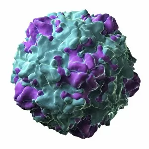
Background imageVirology Collection: Flock House virus, artwork

Flock House virus, artwork
Flock House virus, computer artwork. This is an insect virus that is also able to replicate in plant and yeast cells

Background imageVirology Collection: Equine rhinitis A virus, artwork

Equine rhinitis A virus, artwork
Equine rhinitis A virus capsid, computer artwork. This virus causes respiratory disease in horses

Background imageVirology Collection: Foot and mouth infection, artwork

Foot and mouth infection, artwork
Foot and mouth infection. Computer artwork of foot and mouth virus particles in the bloodstream

Background imageVirology Collection: Echovirus particle, artwork

Echovirus particle, artwork
Echovirus particle, computer artwork. This common human pathogen infects the gastrointestinal tract

Background imageVirology Collection: HPV infection, artwork

HPV infection, artwork
HPV infection. Computer artwork of human papillomavirus particles in the bloodstream

Background imageVirology Collection: Melon necrotic spot virus, artwork

Melon necrotic spot virus, artwork
Melon necrotic spot virus, computer artwork

Background imageVirology Collection: Feline calicivirus, artwork

Feline calicivirus, artwork
Feline calicivirus, computer artwork. This virus causes glossitis (inflammation of the tongue), conjunctivitis (inflammation of the covering of the eye)

Background imageVirology Collection: Swine vesicular disease virus particle F005 / 0712

Swine vesicular disease virus particle F005 / 0712
Swine vesicular disease virus particle, computer model. This virus causes fever and mouth and skin ulcers in pigs

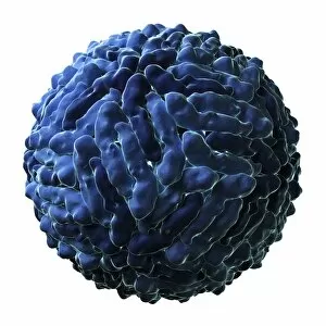
Background imageVirology Collection: Dengue virus particle F005 / 0688

Dengue virus particle F005 / 0688
Dengue virus particle, computer model

Background imageVirology Collection: Hepatitis B virus particle F005 / 0700

Hepatitis B virus particle F005 / 0700
Hepatitis B virus particle, computer model. This virus causes hepatitis, an inflammation of the liver that can lead to cirrhosis and cancer


All Professionally Made to Order for Quick Shipping

Virology, the fascinating study of viruses and their impact on living organisms, delves into a vast array of viral entities that have shaped our understanding of infectious diseases. From the Avian flu virus to HIV particles, Norovirus particles observed through TEM, and even the intricate workings of HIV reverse transcription enzyme and Hepatitis C virus enzyme molecular models - virologists explore these tiny but powerful agents with great curiosity. The Avian flu virus has long been a concern due to its potential for devastating outbreaks in both avian species and humans. Its ability to jump from birds to humans highlights the ever-present threat posed by zoonotic infections. Similarly, HIV particles continue to challenge researchers as they strive for effective treatments against this relentless retrovirus. Norovirus particles captured under transmission electron microscopy (TEM) reveal their unique structure responsible for causing gastroenteritis outbreaks worldwide. Understanding these minute details aids in developing strategies to combat their spread. Hepatitis B viruses are another focus within virology research due to their significant impact on liver health globally. Molecular models help visualize the complex mechanisms employed by these viruses during infection. Microscopic views of human respiratory syncytial virus remind us of how easily respiratory infections can spread among individuals through coughing or sneezing. Artwork depicting such scenarios serves as a reminder of the importance of hygiene practices in preventing disease transmission. Flu virus particle artwork showcases its distinct appearance while emphasizing its continuous evolution and ability to cause seasonal epidemics worldwide. This visual representation helps scientists comprehend its structure better when designing vaccines each year. Coronavirus particles imaged using TEM highlight recent concerns surrounding emerging viral threats like SARS-CoV-2, which causes COVID-19. The ongoing pandemic underscores the urgency for further virological studies aimed at combating future outbreaks effectively. Lastly, adenoviruses capture attention with artistic renditions showcasing their structural intricacies while reminding us that not all viruses are harmful.