
Virus Collection (page 17)
"Unseen Threats: Exploring the Intricacies of Viruses and their Impact on Humanity" Avian flu virus: A microscopic view of the avian flu virus


All Professionally Made to Order for Quick Shipping
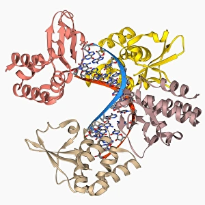
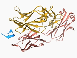
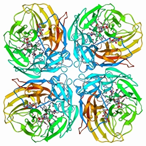
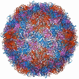
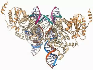
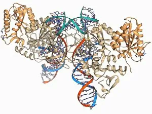
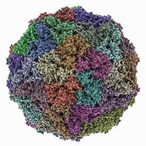
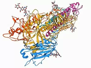
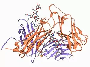
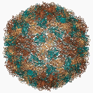
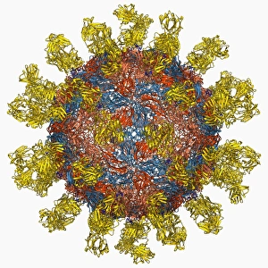
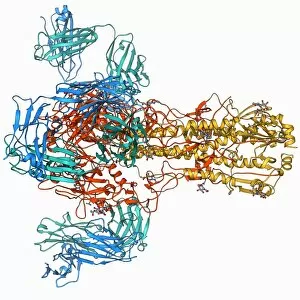
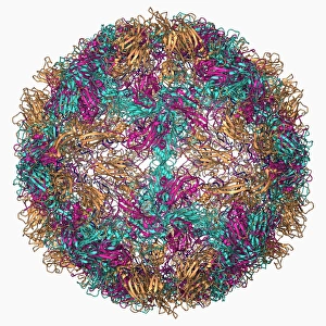
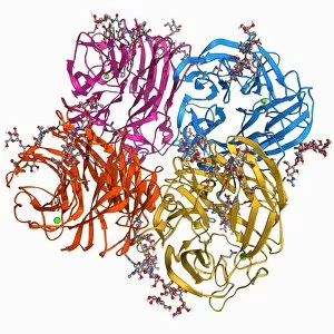
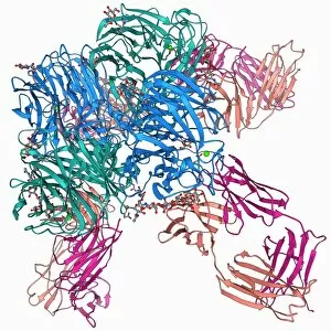
"Unseen Threats: Exploring the Intricacies of Viruses and their Impact on Humanity" Avian flu virus: A microscopic view of the avian flu virus, a notorious pathogen that poses a significant threat to both birds and humans. Unveiling HIV: Delving into the intricate structure of an HIV particle, shedding light on one of the most challenging viruses humanity has faced. Norovirus Particles Revealed: Captured through a powerful TEM microscope, these norovirus particles showcase their unique shape and potential for causing severe gastrointestinal illness. Decoding Hepatitis C: A molecular model displays the complex enzyme responsible for Hepatitis C infection, highlighting ongoing efforts to combat this silent killer. Alexandre Yersin's Discovery: Paying homage to Alexandre Yersin, whose groundbreaking research led to identifying the causative agent behind bubonic plague - Yersinia pestis bacteria. Avian Flu Resurgence: With avian flu outbreaks resurfacing globally, understanding its transmission dynamics becomes crucial in preventing future pandemics. Battling Hepatitis B: Molecular models depict hepatitis B viruses - a major cause of liver disease worldwide - emphasizing the importance of vaccination campaigns and public health initiatives. Empowering Indigenous Communities against Viral Threats in Mexico: Highlighting efforts made towards educating indigenous populations about viral diseases amidst poverty-stricken conditions in Mexico. Coronavirus Artwork Reflects Global Crisis: An artistic representation captures the essence of coronavirus' impact on society, reminding us all to remain vigilant in our fight against this relentless enemy. Broken Tulip Syndrome Exposed: The example of a broken tulip serves as an analogy for how certain plant viruses can devastate entire crops, posing economic challenges for farmers worldwide. Respiratory Syncytial Virus Under Microscope Lens.