
Viscera Collection
"Exploring the Intricate World of Viscera: From Pig Anatomy to Canopic Jars" Delving into the depths of anatomical wonders


All Professionally Made to Order for Quick Shipping
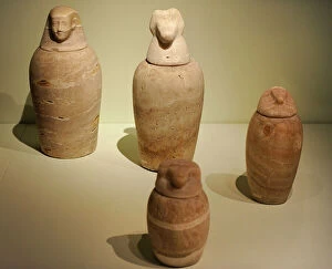
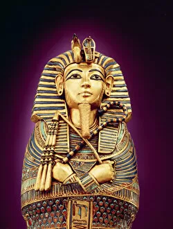
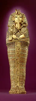
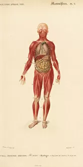
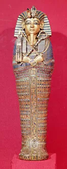
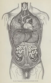
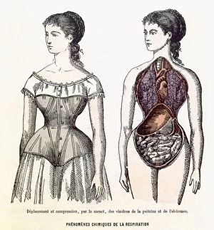
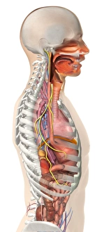
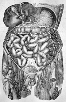
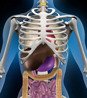
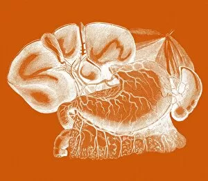
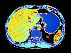
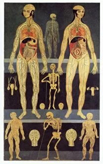
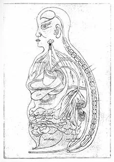
"Exploring the Intricate World of Viscera: From Pig Anatomy to Canopic Jars" Delving into the depths of anatomical wonders, one cannot ignore the intricate world of viscera. Whether it be through pig anatomy or mesmerizing artwork, this captivating subject unveils a multitude of secrets. In ancient Egypt, canopic jars were used to preserve organs during mummification. These beautifully crafted vessels held the key to eternal life as they safeguarded essential parts such as lungs and other viscera. The interior of one such canopic coffin discovered in Tutankhamun's tomb reveals its opulence with beaten gold adorning every inch. The attention to detail is awe-inspiring as we observe the front view of these canopic coffins. Each intricately carved feature showcases not only artistic prowess but also reverence for preserving life beyond death. Rear view exposes further mysteries concealed within these sacred containers. Moving away from ancient Egypt, our exploration takes us on a journey through various animal anatomies. The thoracic viscera of horses provides insight into their internal workings while lithographs depict an exquisite portrayal of their musculature and internal organs. Even smaller creatures like frogs are not exempt from our study; opened up, they reveal delicate lungs and other vital viscera that keep them alive in their watery habitats. Such discoveries remind us that even in nature's tiniest creations, complexity thrives. Lastly, human musculature and internal organs offer an introspective look at ourselves - a reminder that beneath our skin lies a network responsible for sustaining life itself, and is through understanding these intricate systems that medical advancements continue to flourish. Viscera captivates both scientific minds and art enthusiasts alike by showcasing the beauty hidden within living organisms' innermost sanctums. From pig anatomy to canopic jars found in Tutankhamun's tomb or even depictions of horse thoracic viscera – each revelation unravels the wonders of life's intricate design.