Fraud Blocker Skip to main content

Visual Sense Collection (page 3)

"Unlocking the Wonders of Visual Sense: Exploring the Intricacies of Our Eyes" Our eyes, like intricate masterpieces, hold within them a world of visual wonders

Background imageVisual Sense Collection: Artwork of the structure of the front of an eye

Artwork of the structure of the front of an eye

Background imageVisual Sense Collection: Coloured SEM of rod cells in the retina of the eye

Coloured SEM of rod cells in the retina of the eye
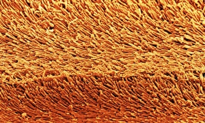
Rod cells. Coloured scanning electron micrograph (SEM) of a group of rod cells in the retina of the eye. Rod cells are light sensitive cells which respond to dim light

Background imageVisual Sense Collection: Coloured SEM of iris epithelial cells of the eye

Coloured SEM of iris epithelial cells of the eye
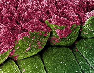
Iris epithelium. Coloured scanning electron micrograph (SEM) of epithelial cells (green) lining the underside of the human iris

Background imageVisual Sense Collection: Eye retina

Eye retina
Structure of the retina. Computer artwork showing the structure of the eye retina. The retina is a thin tissue layer on the inner eye responsible for sight. Light strikes from the top

Background imageVisual Sense Collection: Computer artwork of the structure of the retina

Computer artwork of the structure of the retina
Structure of the retina. Computer artwork showing the structure of the eye retina. The retina is a thin tissue layer on the inner eye responsible for sight. Light strikes from the top

Background imageVisual Sense Collection: Blood vessels in eye

Blood vessels in eye
False-colour scanning electron micrograph (SEM) of blood vessels in the choroid of the eye. A branch- ing network of arteries and veins can be seen in this area under the central fovea

Background imageVisual Sense Collection: False-colour SEM of the structure of the retina

False-colour SEM of the structure of the retina
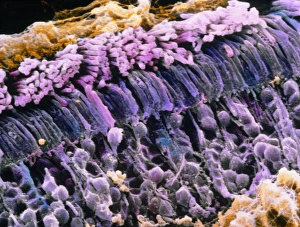
False-colour scanning electron micrograph (SEM) of the eye retina. The retina, which detects visible images, is made up of many layers of cells (seen here)

Background imageVisual Sense Collection: False-colour SEM of rod cells in human retina

False-colour SEM of rod cells in human retina
False-colour scanning electron micrograph (SEM) of rod cells situated in the human retina. Two layers are visible here : one at top (pinkish)

Background imageVisual Sense Collection: False-colour SEM of cells from lens of hu

False-colour SEM of cells from lens of hu

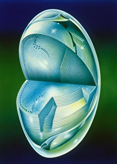
Background imageVisual Sense Collection: Illustration of the fine structure of the eye lens

Illustration of the fine structure of the eye lens. This is the internal optical component (also called the crystalline lens) of the eye, responsible for adjusting focus

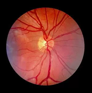
Background imageVisual Sense Collection: Normal retina of eye

Normal retina of eye
Fundus camera image of the retina of a normal eye, showing the distribution of the retinal veins & arteries: the central retinal artery (a branch of the opthalmic artery)

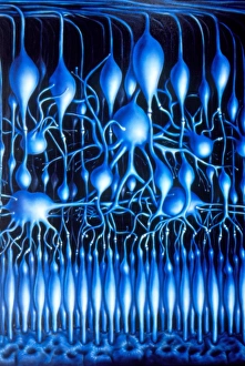
Background imageVisual Sense Collection: Illustration of nerve cells in mammalian retina

Illustration of nerve cells in mammalian retina
Illustration of the layers of nerve cells comprising the mammalian retina, the light- sensitive layer of the eye. At bottom

Background imageVisual Sense Collection: Contact lens

Contact lens
MODEL RELEASED. Contact lens. Woman inserting a disposable (soft) contact lens

Background imageVisual Sense Collection: Magnified eye

Magnified eye
MODEL RELEASED. Magnified eye seen through a magnifying glass

Background imageVisual Sense Collection: Eye

Eye. Computer artwork of a female eye with a maze superimposed on the iris (green)

Background imageVisual Sense Collection: Human eye

Human eye, computer artwork

Background imageVisual Sense Collection: Vision

Vision. Glowing human eye

Background imageVisual Sense Collection: Eye

Eye. Computer artwork of a close-up of the iris and pupil of an eye. The iris, a coloured muscular ring, regulates the amount of light that enters the eye through the pupil (black)

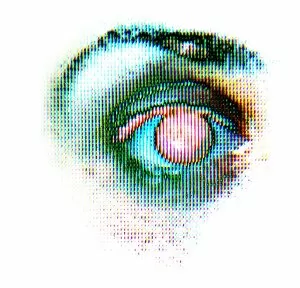
Background imageVisual Sense Collection: Pixellated eye

Pixellated eye, computer artwork. This could represent artificial vision or impaired sight

Background imageVisual Sense Collection: Eye

Eye. Close-up of a womans eye

Background imageVisual Sense Collection: Inserting contact lens

Inserting contact lens
Contact lens. Artwork of a woman putting in a con- tact lens. Contact lenses alter the focusing power of the cornea, the transparent front part of the eye that focuses light onto the back of the eye

Background imageVisual Sense Collection: Womans eye

Womans eye
Womans blue eye

Background imageVisual Sense Collection: Biometrics, conceptual artwork

Biometrics, conceptual artwork
MODEL RELEASED. Biometrics, conceptual computer artwork

Background imageVisual Sense Collection: Laser eye surgery, computer artwork

Laser eye surgery, computer artwork
Computer artwork depicting laser eye surgery. Lasers have a variety of uses in ophthalmic surgery, including photo-cauterisation of blood vessels in retinopathy

Background imageVisual Sense Collection: Retina blood vessels and nerve cells

Retina blood vessels and nerve cells. Light micrograph showing the blood vessels and nerve cells in the retina of an eye. The retina is the light-sensitive membrane that lines the back of the eyeball

Background imageVisual Sense Collection: Visual pathways, artwork

Visual pathways, artwork
Visual pathways. Artwork of the visual pathways in the human brain. The optic nerves (white and blue lines) extend from the retinas at the back of each eye towards the optic chiasma

Background imageVisual Sense Collection: Mans eye

Mans eye
MODEL RELEASED. Mans eye. Close up of a healthy mans eye

Background imageVisual Sense Collection: Eye lens fibres, SEM

Eye lens fibres, SEM
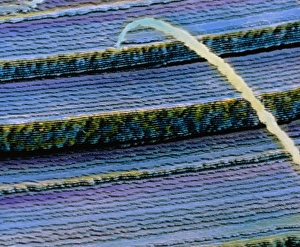
Eye lens fibres. Coloured scanning electron micrograph (SEM) of a freeze-fracture of a lens of an eye, showing the regular arrangement of fibres


All Professionally Made to Order for Quick Shipping

"Unlocking the Wonders of Visual Sense: Exploring the Intricacies of Our Eyes" Our eyes, like intricate masterpieces, hold within them a world of visual wonders. The iris, akin to a delicate flower's petals, reveals its captivating hues and patterns that make each individual unique. Through the lens of a scanning electron microscope (SEM), we delve into the mesmerizing details of this remarkable feature. As we journey deeper into our visual sense, contact lenses emerge as modern marvels that enhance our perception. They seamlessly blend with our irises, offering clarity and comfort while allowing us to see the world in all its vibrant glory. The retina takes center stage in this symphony of sight – an intricately woven tapestry capturing light and transforming it into vivid images. Its false-color SEM unveils the central fovea, where vision is sharpest and most precise; a testament to nature's precision craftsmanship. Delving further into this microscopic realm, we encounter the ciliary body – an unsung hero responsible for adjusting our eye's focus effortlessly. Revealed by SEM imaging techniques, its intricate structures showcase nature's ingenuity at work. Traversing through retinal blood vessels captured by SEM imagery unravels their complex network nourishing every corner of our visual canvas. These lifelines remind us how interconnected every aspect of our visual sense truly is. The eye lens emerges as another extraordinary component under scrutiny by SEM technology - revealing its layered architecture designed meticulously for optimal refraction and focusing power. It serves as a gateway between worlds - bending light rays to create clear visions before they reach our precious retina. Within this masterpiece lies rods and cones - tiny warriors guarding against darkness or illuminating colors with finesse unmatched elsewhere in nature’s palette. Their artistic portrayal showcases their vital role in shaping what we perceive around us. Lastly but not leastly comes eye color – an enchanting kaleidoscope reflecting both genetics and individuality.