
Windpipe Collection
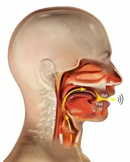
The windpipe, also known as the trachea, is a vital component of the respiratory system. It serves as a passageway for air to travel from the throat and into the lungs


All Professionally Made to Order for Quick Shipping
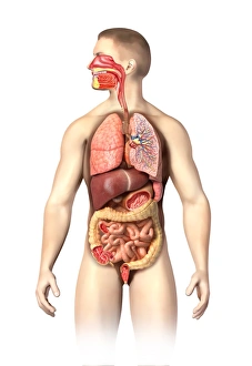
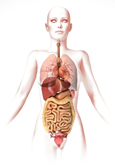
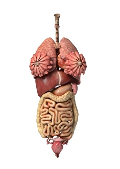
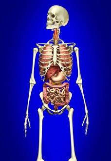
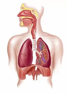
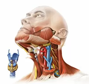
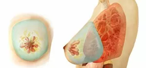
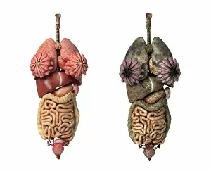
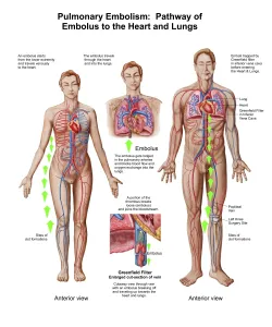
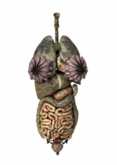
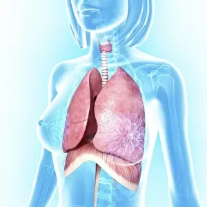
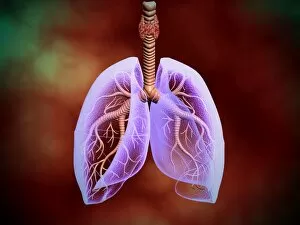
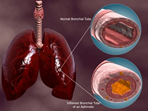
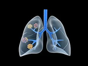
The windpipe, also known as the trachea, is a vital component of the respiratory system. It serves as a passageway for air to travel from the throat and into the lungs. The anatomy of the bronchus and bronchial tubes can be seen in intricate detail within this complex organ. In both male and female bodies, the windpipe is situated amidst a network of internal organs that work together to facilitate respiration. The male respiratory system showcases its connection with other vital structures such as the throat and jaw, which are beautifully illustrated in Manuel d'Anatomie descriptive du Corps. Tracheotomy operations have been extensively studied throughout medical history. Plates from Traite Complet de l'Anatomie de l'Homme provide visual insights into this surgical procedure, highlighting how incisions are made in order to access or bypass obstructions within the windpipe. The importance of understanding these anatomical intricacies becomes evident when considering conditions affecting the respiratory system. For instance, Atlas and Epitome of Operative Surgery by Dr depicts various techniques used during tracheotomy procedures to alleviate breathing difficulties caused by blockages or injuries. Furthermore, an appreciation for how internal organs interact with one another is crucial when examining disorders related to both respiration and circulation. Plates from Anatomy of the Visceras dissected demonstrate how interconnected systems like the throat and heart coexist harmoniously within our bodies. Manuel d'Anatomie descriptive du Corps further enhances our knowledge by providing comprehensive illustrations depicting not only specific regions but also broader perspectives on organ placement within chest cavities. Lastly, advancements in technology have allowed us to explore human anatomy through 3D renderings. These cutting-edge visuals enable us to visualize healthy female internal organs accurately while appreciating their complexity at a whole new level. Studying different aspects surrounding windpipe anatomy provides invaluable insights into its role within our bodies.