
X Ray Image Collection
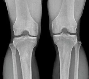
"Unveiling the Invisible: A Glimpse into the Fascinating World of X-ray Images" "Beneath the Surface: Exploring Normal Knees through X-ray Vision" "Peering Inside


All Professionally Made to Order for Quick Shipping
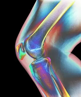
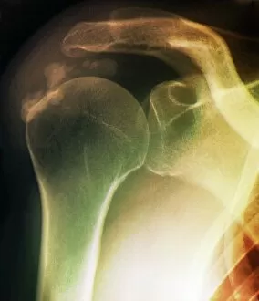
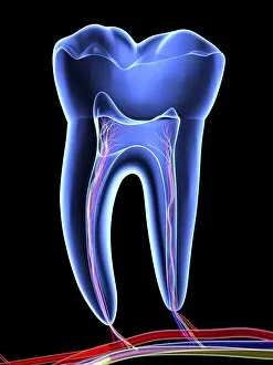
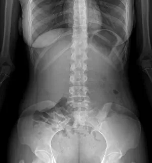
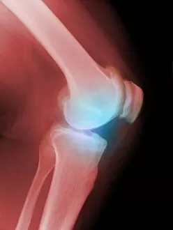
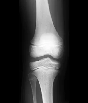
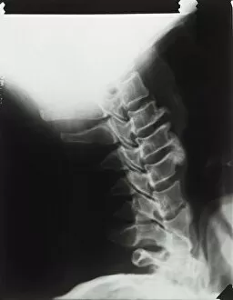
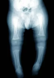
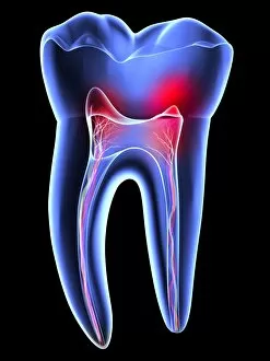
"Unveiling the Invisible: A Glimpse into the Fascinating World of X-ray Images" "Beneath the Surface: Exploring Normal Knees through X-ray Vision" "Peering Inside: The Astonishing Clarity of a Normal Knee in an X-ray Image" "Shoulder Troubles Revealed: Tendinitis Unmasked by an X-ray Scan" "Innocence Preserved: Discovering a Normal Child's Head through an X-ray Lens" "X-Ray Fashionista: Rocking the 'X-Ray Outfit' with Style and Confidence. " "Beyond Waves, Beyond Horizons: Cruising Through a Ship's Inner Secrets via X-rays" "Tooth Tales Unveiled: Journeying into the Depths of Molar Mysteries with an X-ray Image" "Aging Joints Exposed: Witnessing Arthritic Knees through Crystal Clear X-rays" "Legs Underneath It All: Appreciating the Beauty of a Normal Lower Leg in an X-ray View" "Mind-Boggling Precision: Peeking into Perfection with a Normal Skull in an X-ray Snapshot" "Inside Story Unfolded. Decoding Abdominal Health with Detailed Insights from an X-ray Image. " "The Battle Within.