Fraud Blocker Skip to main content

X Rays Collection

"Unveiling the Hidden: Exploring the World of X-Rays" Step into a world where secrets are revealed and mysteries unfold

Background imageX Rays Collection: Skeletons, X-ray artwork

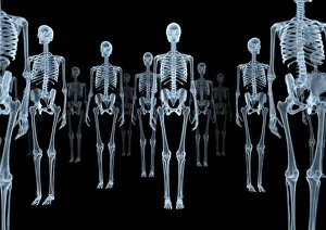
Skeletons, X-ray artwork
Skeleton crowd. Computer X-ray artwork of a group of human skeletons viewed from the front

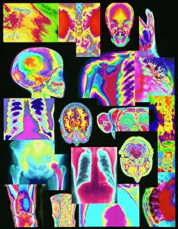
Background imageX Rays Collection: Assortment of coloured X-rays and body scans

Assortment of coloured X-rays and body scans
X-ray diagnosis. Assortment of coloured X-rays, body and brain scans

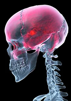
Background imageX Rays Collection: Headache, X-ray artwork

Headache, X-ray artwork
Headache. Computer X-ray artwork of a lateral view of a human skull. The red area represents pain caused by a headache (cephalalgia)

Background imageX Rays Collection: Shells - x-ray

Shells - x-ray
PM-7161 SHELLS - X-RAY Pat Morris Please note that prints are for personal display purposes only and may not be reproduced in any way. contact details: prints@ardea.com tel: + 44 (0) 20 8318 1401

Background imageX Rays Collection: Skeleton from below, X-ray artwork

Skeleton from below, X-ray artwork
Skeleton. Computer x-ray artwork of a human skeleton viewed from below

Background imageX Rays Collection: X-Ray - Salamander

X-Ray - Salamander. Part of Box 165 Boswell Collection - X-Rays. Date: circa 1900

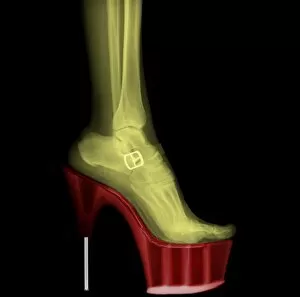
Background imageX Rays Collection: X-ray Stiletto High-Heeled Shoe

X-ray Stiletto High-Heeled Shoe
Coloured X-ray of a female leg wearing Stiletto High-Heeled Shoe

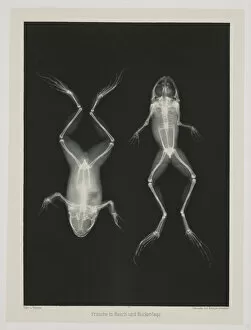
Background imageX Rays Collection: Diagnostic Radiology, X-ray of two frogs, 1896

Diagnostic Radiology, X-ray of two frogs, 1896
UIG5086339 Diagnostic Radiology, X-ray of two frogs, 1896 by Eder, Josef Maria (1855 - 1944); (add.info.: Diagnostic Radiology, X-rays X-ray of two frogs, 1896)

Background imageX Rays Collection: X-ray security machine, 1900

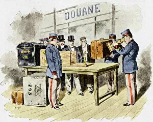
X-ray security machine, 1900
X-ray security machine. Historical artwork of a fluoroscope being used in a late 19th-century French customs hall

Background imageX Rays Collection: Electromagnetic spectrum and its visible spectrum highlighted separately

Electromagnetic spectrum and its visible spectrum highlighted separately

Background imageX Rays Collection: Penetrating power of Alpha, Beta and Gamma ray through Paper

Penetrating power of Alpha, Beta and Gamma ray through Paper, Aluminium, Led and Concrete. Penetration power of alpha, beta and gamma radiation

Background imageX Rays Collection: Dentist prepares smiling girl wearing in protective lead apron to jaw x-ray image in dental clinic

Dentist prepares smiling girl wearing in protective lead apron to jaw x-ray image in dental clinic. Focus on woman

Background imageX Rays Collection: Solus-Schall Industrial Radiography. NDT X-Rays Gamma Rays

Solus-Schall Industrial Radiography. NDT X-Rays Gamma Rays - Solus-Schall Ltd. Date: 1952

Background imageX Rays Collection: Solus-Schall Industrial Radiography. NDT X-Rays Gamma Rays

Solus-Schall Industrial Radiography. NDT X-Rays Gamma Rays - Solus-Schall Ltd. Date: 1952

Background imageX Rays Collection: Solus-Schall Industrial Radiography. NDT X-Rays Gamma Rays

Solus-Schall Industrial Radiography. NDT X-Rays Gamma Rays - Solus-Schall Ltd. Date: 1952

Background imageX Rays Collection: Solus-Schall Industrial Radiography. NDT X-Rays Gamma Rays

Solus-Schall Industrial Radiography. NDT X-Rays Gamma Rays - Solus-Schall Ltd. Date: 1952

Background imageX Rays Collection: Normal legs, X-rays

Normal legs, X-rays
Normal legs. Coloured frontal X-rays of the healthy legs, of a 35-year-old

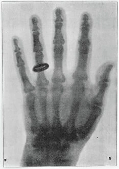
Background imageX Rays Collection: X-ray of the left hand of Anna Bertha Ludwig, Rontgen's wife, 1895 (b/w photo)

X-ray of the left hand of Anna Bertha Ludwig, Rontgen's wife, 1895 (b/w photo)
CHT207897 X-ray of the left hand of Anna Bertha Ludwig, Rontgen's wife, 1895 (b/w photo) by Rontgen

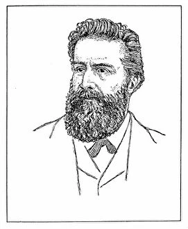
Background imageX Rays Collection: Portrait of William Konrad Von Rontgen, 1845-1923 (litho)

Portrait of William Konrad Von Rontgen, 1845-1923 (litho)
8634687 Portrait of William Konrad Von Rontgen, 1845-1923 (litho) by English School, (20th century); Private Collection; (add.info.: William Konrad Von Rontgen, 1845-1923)

Background imageX Rays Collection: Letter X: X-ray (X-ray). Little French alphabet

Letter X: X-ray (X-ray). Little French alphabet
DUV4204721 Letter X: X-ray (X-ray). Little French alphabet. Hachette and Cie, publisher, Paris, 1917 (lithograph); 30.5x23.5 cm; Private Collection; (add.info.: Illustrations by J. M. Porta. 8 pages)

Background imageX Rays Collection: Letter W and X: Watman and X-ray (X-ray)

Letter W and X: Watman and X-ray (X-ray)
DUV4204713 Letter W and X: Watman and X-ray (X-ray). Little French alphabet. Hachette and Cie, publisher, Paris, 1917 (lithograph); 30.5x23.5 cm; Private Collection; (add.info.: Illustrations by J)

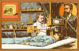
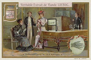
Background imageX Rays Collection: X-rays and radiography. Position of needle in woman's hand being shown being

X-rays and radiography. Position of needle in woman's hand being shown being
527525 X-rays and radiography. Position of needle in woman's hand being shown being. From Liebig trade card c.1900. Chromolithograph ; Universal History Archive/UIG

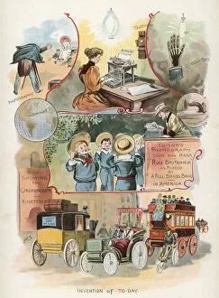
Background imageX Rays Collection: Transport and technology in 1875 and 1900 (chromolitho)

Transport and technology in 1875 and 1900 (chromolitho)
8677571 Transport and technology in 1875 and 1900 (chromolitho) by American School

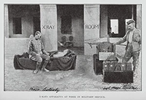
Background imageX Rays Collection: X-ray Apparatus at Work in Military Service (b/w photo)

X-ray Apparatus at Work in Military Service (b/w photo)
8639575 X-ray Apparatus at Work in Military Service (b/w photo) by English Photographer, (20th century); Private Collection; (add.info.: X-ray Apparatus at Work in Military Service)

Background imageX Rays Collection: This Mechanized Age: X-ray apparatus (colour photo)

This Mechanized Age: X-ray apparatus (colour photo)
8660468 This Mechanized Age: X-ray apparatus (colour photo) by English Photographer, (20th century); Private Collection; (add.info.: X-ray apparatus)

Background imageX Rays Collection: X-ray - Soldier with Bullet Bound Being Examined 1900

X-ray - Soldier with Bullet Bound Being Examined 1900
Wounded soldier from the Boer War been examined in a London hospital by means of X-ray. One of the interesting features of the Boer War was the demonstration of the practical utility of the X-rays

Background imageX Rays Collection: Ladysmith Hospital Corps (b / w photo)

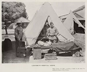
Ladysmith Hospital Corps (b / w photo)
7160180 Ladysmith Hospital Corps (b/w photo) by Kisch, David Henry (1847-1902); Private Collection; (add.info.: Ladysmith Hospital Corps)

Background imageX Rays Collection: Portrait of Wilhelm Rontgen, 1915 (photo)

Portrait of Wilhelm Rontgen, 1915 (photo)
3433059 Portrait of Wilhelm Rontgen, 1915 (photo) by Perscheid

Background imageX Rays Collection: Controlling powers of destruction (litho)

Controlling powers of destruction (litho)
979570 Controlling powers of destruction (litho) by Begg, Samuel (fl.1886-1916) (after); Private Collection; (add.info.: Controlling powers of destruction)

Background imageX Rays Collection: X-Rays (chromolitho)

X-Rays (chromolitho)
3643479 X-Rays (chromolitho) by French School, (19th century); Private Collection; (add.info.: X-Rays. Educational card, late 19th or early 20th century.); © Look and Learn

Background imageX Rays Collection: Inventions

Inventions
LLM462053 Inventions by Sinclair, J. R. (19th century); Private Collection; (add.info.: Inventions. Illustration for The Victorian Picture Book by W H Stacpoole (Dean, c 1900)); © Look and Learn

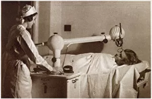
Background imageX Rays Collection: Apparatus that Assist the Remedies of Nature 1933

Apparatus that Assist the Remedies of Nature 1933
A cure for facial blemishes: The Siemens-Reinger apparatus, treatment by the means of X-rays anything from tiny warl to a widespread skin disease. Date: 1933

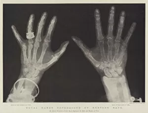
Background imageX Rays Collection: Royal Hands reproduced by Rontgen Rays (engraving)

Royal Hands reproduced by Rontgen Rays (engraving)
1605592 Royal Hands reproduced by Rontgen Rays (engraving) by English School, (19th century); Private Collection; (add.info.: Royal Hands reproduced by Rontgen Rays)

Background imageX Rays Collection: X-ray had proven useful in World War One, for locating bullets

X-ray had proven useful in World War One, for locating bullets and foreign bodies in wounded men and also detection of any contraband

Background imageX Rays Collection: Man having a chest X-ray (b / w photo)

Man having a chest X-ray (b / w photo)
6015600 Man having a chest X-ray (b/w photo) by English Photographer, (20th century); Private Collection; (add.info.: Man having a chest X-ray)

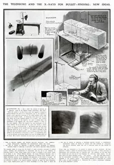
Background imageX Rays Collection: New Ideas for using X-Rays for Medical use WWI

New Ideas for using X-Rays for Medical use WWI
Used in the British, French and Russian military hospitals the Kompact, apparatus, to find foreign bodies in wounded soldiers

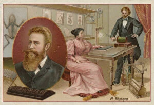
Background imageX Rays Collection: Wilhelm Roentgen, German physicist who discovered X-rays (chromolitho)

Wilhelm Roentgen, German physicist who discovered X-rays (chromolitho)
972094 Wilhelm Roentgen, German physicist who discovered X-rays (chromolitho) by French School, (19th century); Private Collection; (add.info.: Wilhelm Roentgen (1845-1923)

Background imageX Rays Collection: X rays - no need to open letters to read them any more (chromolitho)

X rays - no need to open letters to read them any more (chromolitho)
1094111 X rays - no need to open letters to read them any more (chromolitho) by French School, (19th century); Private Collection; (add.info.: X rays - no need to open letters to read them any more)

Background imageX Rays Collection: Radiography and X-rays (chromolitho)

Radiography and X-rays (chromolitho)
974602 Radiography and X-rays (chromolitho) by European School, (19th century); Private Collection; (add.info.: Radiography and X-rays)

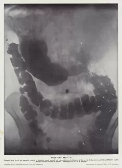
Background imageX Rays Collection: Roentgen Rays, II (b / w photo)

Roentgen Rays, II (b / w photo)
3624628 Roentgen Rays, II (b/w photo) by English Photographer, (20th century); Private Collection; (add.info.: Roentgen Rays, II)

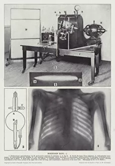
Background imageX Rays Collection: Roentgen Rays, I (b / w photo)

Roentgen Rays, I (b / w photo)
3624627 Roentgen Rays, I (b/w photo) by English Photographer, (20th century); Private Collection; (add.info.: Roentgen Rays, I)

Background imageX Rays Collection: Beach idyll in Rontgen style, cartoon about X-rays, c. 1896-99 (colour engraving)

Beach idyll in Rontgen style, cartoon about X-rays, c. 1896-99 (colour engraving)
CHT164182 Beach idyll in Rontgen style, cartoon about X-rays, c.1896-99 (colour engraving) by German School, (19th century); Bibliotheque des Arts Decoratifs, Paris

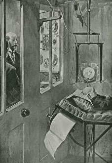
Background imageX Rays Collection: Finding a bullet with the X-ray apparatus at the Military Hospital

Finding a bullet with the X-ray apparatus at the Military Hospital
5236263 Finding a bullet with the X-ray apparatus at the Military Hospital, Pietermaritzburg, Natal, South Africa, Boer War, 1899 (litho) by Bull

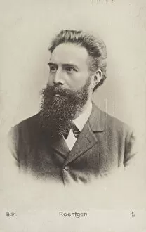
Background imageX Rays Collection: Wilhelm Roentgen (1845-1923), German physicist (b / w photo)

Wilhelm Roentgen (1845-1923), German physicist (b / w photo)
817385 Wilhelm Roentgen (1845-1923), German physicist (b/w photo) by German Photographer, (19th century); Private Collection; (add.info.: Wilhelm Roentgen (1845-1923)

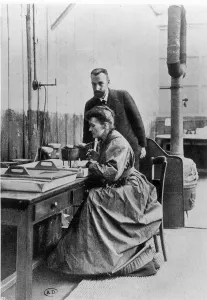
Background imageX Rays Collection: Pierre (1859-1906) and Marie Curie (1867-1934) in their laboratory, c. 1903 (b / w photo)

Pierre (1859-1906) and Marie Curie (1867-1934) in their laboratory, c. 1903 (b / w photo)
CHT237251 Pierre (1859-1906) and Marie Curie (1867-1934) in their laboratory, c.1903 (b/w photo) by French Photographer, (20th century); Bibliotheque des Arts Decoratifs, Paris

Background imageX Rays Collection: Pierre Curie (1859-1906) (b / w photo)

Pierre Curie (1859-1906) (b / w photo)
ACD237252 Pierre Curie (1859-1906) (b/w photo) by French Photographer, (20th century); Academie des Sciences, Paris

Background imageX Rays Collection: Wilhelm Roentgen, German physicist (chromolitho)

Wilhelm Roentgen, German physicist (chromolitho)
972119 Wilhelm Roentgen, German physicist (chromolitho) by Swiss School, (19th Century); Private Collection; (add.info.: Wilhelm Roentgen (1845-1923), German physicist who discovered X-rays

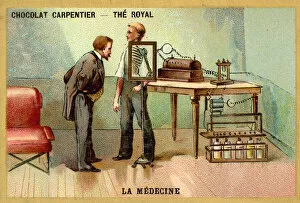
Background imageX Rays Collection: Patient having an x-ray (chromolitho)

Patient having an x-ray (chromolitho)
1093981 Patient having an x-ray (chromolitho) by French School, (19th century); Private Collection; (add.info.: Patient having an x-ray)

Background imageX Rays Collection: Wilhelm Roentgen, German physicist who discovered X-rays. (b / w photo)

Wilhelm Roentgen, German physicist who discovered X-rays. (b / w photo)
6014940 Wilhelm Roentgen, German physicist who discovered X-rays

Background imageX Rays Collection: Wilhelm Roentgen (1845-1923), German physicist (b / w photo)

Wilhelm Roentgen (1845-1923), German physicist (b / w photo)
817386 Wilhelm Roentgen (1845-1923), German physicist (b/w photo) by German Photographer, (19th century); Private Collection; (add.info.: Wilhelm Roentgen (1845-1923)

Background imageX Rays Collection: The possibilities of the Rontgen Method (engraving)

The possibilities of the Rontgen Method (engraving)
3621272 The possibilities of the Rontgen Method (engraving) by English School, (19th century); Private Collection; (add.info.: The possibilities of the Rontgen Method)

Background imageX Rays Collection: Advertisement for Philips X-Ray Shoe Fitter, 1930s (litho)

Advertisement for Philips X-Ray Shoe Fitter, 1930s (litho)
736244 Advertisement for Philips X-Ray Shoe Fitter, 1930s (litho) by English School, (20th century); Private Collection; (add.info.: Advertisement for Philips X-Ray Shoe Fitter)

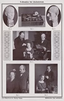
Background imageX Rays Collection: Personality analysis using x-rays (b / w photo)

Personality analysis using x-rays (b / w photo)
6000728 Personality analysis using x-rays (b/w photo) by German Photographer (20th Century); Private Collection; (add.info.: Personality analysis using x-rays)

Background imageX Rays Collection: X-rays (chromolitho)

X-rays (chromolitho)
972126 X-rays (chromolitho) by French School, (19th century); Private Collection; (add.info.: X-rays. Educational card.); © Look and Learn

Background imageX Rays Collection: The Bones of a Hand photographed through the Flesh by means of the New Process (litho)

The Bones of a Hand photographed through the Flesh by means of the New Process (litho)
1041902 The Bones of a Hand photographed through the Flesh by means of the New Process (litho) by English School

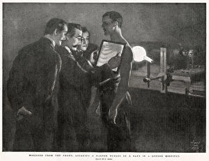
Background imageX Rays Collection: Wounded from the Front, locating a Mauser Bullet by X Rays in a London Hospital (engraving)

Wounded from the Front, locating a Mauser Bullet by X Rays in a London Hospital (engraving)
1048542 Wounded from the Front, locating a Mauser Bullet by X Rays in a London Hospital (engraving) by Small, William (1843-1931) (after); Private Collection; (add.info.: Wounded from the Front)

Background imageX Rays Collection: X-Ray - Elbow Joint

X-Ray - Elbow Joint. Part of Box 165 Boswell Collection - X-Rays. Date: circa 1900

Background imageX Rays Collection: X-Ray - Guinea Pig

X-Ray - Guinea Pig. Part of Box 165 Boswell Collection - X-Rays. Date: circa 1900

Background imageX Rays Collection: X-Ray - Foot of Lamb

X-Ray - Foot of Lamb. Part of Box 165 Boswell Collection - X-Rays. Date: circa 1900

Background imageX Rays Collection: X-Ray - Claw Foot

X-Ray - Claw Foot. Part of Box 165 Boswell Collection - X-Rays. Date: circa 1900

Background imageX Rays Collection: Rontgen, Wilhelm Conrad (1845-1923). German physicist. Rontg

Rontgen, Wilhelm Conrad (1845-1923). German physicist. Rontg
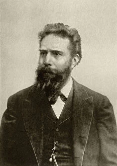
Rontgen, Wilhelm Conrad (1845-1923). German physicist. Discovered Roentgen rays or X-ray (1895). In 1901 he was awarded the Nobel Prize in Physics

Background imageX Rays Collection: Salivary gland stone, X-ray

Salivary gland stone, X-ray
Salivary gland stone. X-ray of the jaw of a 36-year-old patient with a stone (white, lower left) in the right submaxillary gland

Background imageX Rays Collection: Caricature of Roentgen and X-rays

Caricature of Roentgen and X-rays. The German experimental physicist Wilhelm Konrad Roentgen (1845-1923) had discovered X-rays in 1895. This artwork shows his skeleton revealed by the use of X-rays

Background imageX Rays Collection: X-Ray - Bank of Green Fern

X-Ray - Bank of Green Fern. Part of Box 165 Boswell Collection - X-Rays. Date: circa 1900

Background imageX Rays Collection: X-Ray - Honey Bee Sting

X-Ray - Honey Bee Sting. Part of Box 165 Boswell Collection - X-Rays. Date: circa 1900

Background imageX Rays Collection: Diagram of the transparency of Earths atmosphere to different types of radiation

Diagram of the transparency of Earths atmosphere to different types of radiation
Diagram illustrating the transparency of Earths atmosphere to different types of radiation

Background imageX Rays Collection: Normal knees, X-rays

Normal knees, X-rays
Normal knees. Coloured frontal X-rays of the healthy knees of a 35-year-old

Background imageX Rays Collection: X-ray of a couple kissing

X-ray of a couple kissing

Background imageX Rays Collection: Thinking man with a lightbulb

Thinking man with a lightbulb
X-ray of a thinking man with a lightbulb

Background imageX Rays Collection: Throat cancer, X-rays

Throat cancer, X-rays
Throat cancer. Coloured X-rays showing the transit of a contrast medium (dense white) through the throat of a 54-year-old patient with an adenocarcinoma (cancer) of the lower oesophagus (gullet)

Background imageX Rays Collection: Adult and child hand X-rays

Adult and child hand X-rays
Adult and child hands. Coloured X-rays of healthy human hands at 3 years (left) and at 20 years. Bones are green with flesh red

Background imageX Rays Collection: Fractured foot, coloured X-ray

Fractured foot, coloured X-ray
Fractured foot. Coloured X-rays of a fractured fifth metatarsal (to the right of each image). The fracture is seen as a small black crack

Background imageX Rays Collection: Healthy skull, coloured X-ray

Healthy skull, coloured X-ray
Healthy human skull. Side view X-ray showing the skull and the outline of the head (black). The bones of the cranium are fused together to form a complete structure that protects the brain

Background imageX Rays Collection: Neck and shoulder pain, X-ray artwork

Neck and shoulder pain, X-ray artwork
Neck and shoulder pain. Computer X-ray artwork of the back view of a human skeletons shoulders, neck and skull. The red area represents neck pain (cervicalgia) and shoulder pain

Background imageX Rays Collection: Early x-ray of a bird

Early x-ray of a bird
In 1895 Roentgen discovered a new kind of radiant energy which he named x-rays. On December 22nd 1895 he produced the first human radiograph of his wifes hand

Background imageX Rays Collection: Baler shell, x-ray to show internal structure and spiralling

Baler shell, x-ray to show internal structure and spiralling
PM-10568 Baler shell, x-ray to show internal structure and spiralling. spirals Pat Morris Please note that prints are for personal display purposes only and may not be reproduced in any way

Background imageX Rays Collection: X-Ray - Ribcage and internal organs of the upper abdomen

X-Ray - Ribcage and internal organs of the upper abdomen. Part of Box 165 Boswell Collection - X-Rays. Date: circa 1900

Background imageX Rays Collection: X-Ray - Microscopic x-ray view of a small head louse

X-Ray - Microscopic x-ray view of a small head louse. Part of Box 165 Boswell Collection - X-Rays. Date: circa 1900

Background imageX Rays Collection: X-Ray - Internal structire of a plant cell

X-Ray - Internal structire of a plant cell. Part of Box 165 Boswell Collection - X-Rays. Date: circa 1900

Background imageX Rays Collection: X-Ray - Ivy

X-Ray - Ivy. Part of Box 165 Boswell Collection - X-Rays. Date: circa 1900

Background imageX Rays Collection: X-Ray - Mistletoe Leaf

X-Ray - Mistletoe Leaf. Part of Box 165 Boswell Collection - X-Rays. Date: circa 1900

Background imageX Rays Collection: X-Ray - Close-up X-ray view of a Blow Fly Proboscis

X-Ray - Close-up X-ray view of a Blow Fly Proboscis. Part of Box 165 Boswell Collection - X-Rays. Date: circa 1900


All products are expertly crafted, using premium materials, tailored to your specifications and promptly shipped

"Unveiling the Hidden: Exploring the World of X-Rays" Step into a world where secrets are revealed and mysteries unfold. X-rays, not just a medical tool, but an artistic marvel that showcases the intricate beauty hidden within our bodies. From skeletons to x-ray artwork, immerse yourself in this captivating journey. Witness the ethereal elegance of a skeleton from below, as its delicate structure is unveiled through an x-ray artwork. Marvel at the assortment of colored x-rays and body scans that paint a vivid picture of our inner workings. Ever wondered what causes that pounding headache? Delve into the mesmerizing realm of x-ray artwork depicting cerebral vasculitis, capturing both pain and artistry in one frame. Venture beyond human anatomy and explore nature's wonders with shells captured through an x-ray lens. Admire their intricate patterns as they reveal their hidden intricacies. Take a peek into fashion's fascination with transparency as you gaze upon an exquisite x-ray stiletto high-heeled shoe. Witness how even fashion can be transformed by scientific innovation. Travel back in time to 1900 when security machines were first introduced - witness history being made with an intriguing glimpse at an early version of an x-ray security machine. Discover the enchanting world beneath our feet as you encounter an awe-inspiring salamander captured through an x-ray lens. Its skeletal structure becomes a work of art itself, showcasing nature's ingenuity. Experience empathy for those who have endured physical trauma as you observe a broken arm encased in plaster cast through an x-ray image. The resilience displayed amidst adversity is truly remarkable. Transport yourself to another era with a poignant depiction from 1900 - witness soldiers examined under bullet-bound circumstances using cutting-edge technology like never before seen - the power of X-rays changing lives on the battlefield. Intriguingly intertwined between science and creativity lies the captivating universe of X-rays – where skeletons become art, and the invisible becomes visible.

© Copyright Media Storehouse, All Rights Reserved 2003 - 2024
Facebook     Pinterest     100% Payment Secure     Reviews IO