Images Dated 10th March 2005
Available as Framed Prints, Photos, Wall Art and Gift Items
Choose from 123 pictures in our Images Dated 10th March 2005 collection for your Wall Art or Photo Gift. Popular choices include Framed Prints, Canvas Prints, Posters and Jigsaw Puzzles. All professionally made for quick delivery.
Abstract
Aerial
Africa
Animals
Architecture
Arts
Asia
Europe
Historic
Humour
Maps and Charts
North America
Oceania
People
Popular Themes
Posters
Religion
Science
Services
South America
Special Days
Sport
Transportation
All Images
> 2005
>> March
>>> 1 Mar 2005
>>> 10 Mar 2005
>>> 11 Mar 2005
>>> 12 Mar 2005
>>> 13 Mar 2005
>>> 14 Mar 2005
>>> 15 Mar 2005
>>> 16 Mar 2005
>>> 17 Mar 2005
>>> 18 Mar 2005
>>> 19 Mar 2005
>>> 2 Mar 2005
>>> 20 Mar 2005
>>> 21 Mar 2005
>>> 22 Mar 2005
>>> 23 Mar 2005
>>> 24 Mar 2005
>>> 25 Mar 2005
>>> 26 Mar 2005
>>> 27 Mar 2005
>>> 28 Mar 2005
>>> 29 Mar 2005
>>> 3 Mar 2005
>>> 30 Mar 2005
>>> 31 Mar 2005
>>> 4 Mar 2005
>>> 5 Mar 2005
>>> 6 Mar 2005
>>> 7 Mar 2005
>>> 8 Mar 2005
>>> 9 Mar 2005
Collections

open-uri20120928-21739-1i3gvvg
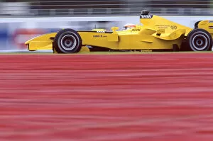
2005 GP2 Series Test.
Ferdinando Monfardini (MC), Durango. IPortrait.
Paul Ricard, France. 8th March 2005.
Photo: GP2 Series Media Service.
Ref: Digital Image Only
© ERDA Communication

Kidney tubules, SEM
Kidney tubules. Coloured scanning electron micrograph (SEM) of proximal convoluted tubules (dark brown) in the kidney. The proximal convoluted tubules function to reabsorb water, glucose and other small molecules from the filtrate, producing a concentrated urine. The tubules consist of a lumen (interior) lined with cuboidal epithelial cells. These cells are covered in many finger-like projections known as microvilli, which serve to increase the cell's surface area for reabsorption. The reabsorbed molecules move into the interstitial fluid of the kidney and are absorbed back into the blood. The liquid in the tubules passes to the bladder. Magnification: x420 when printed 10cm wide
© STEVE GSCHMEISSNER/SCIENCE PHOTO LIBRARY

Artwork of bones & ligaments in human knee joint
Knee joint. Illustration showing bones and tendons of the human knee (genual) joint. The knee is the largest joint in the human body. It forms the point of articulation between the upper leg bone (femur, at top) and the two bones of the lower limb (tibia at lower left, fibula at lower right). The articulation surface is formed from condyles (round bone ends) between the femur and tibia. These condyles are covered in cartilage (white) to prevent friction on movement. Ligaments (pink) fix this joint together: a fibular collateral ligament attaches the fibula to femur; cruciate ligaments attach tibia to femur between the condyles. The oval knee cap bone (patella) protects the joint
© JOHN BAVOSI/SCIENCE PHOTO LIBRARY