Fraud Blocker Skip to main content

Home > All Images > 2007 > October > 31 Oct 2007

Images Dated 31st October 2007 (page 5)

376 items

Background imageImages Dated 31st October 2007: Prostate gland, light micrograph

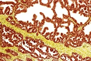
Prostate gland, light micrograph
Prostate gland. Coloured light micrograph of a section through a prostate gland. The prostate is a large male gland that surrounds the neck of the bladder and the urethra

Background imageImages Dated 31st October 2007: Thyroid gland, light micrograph

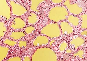
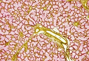
Thyroid gland, light micrograph
Thyroid gland. Coloured light micrograph of a thyroid gland showing the follicles. The follicles are lined by a single layer of cuboidal epithelial cells (purple, dark nuclei)

Background imageImages Dated 31st October 2007: Thyroid gland, light micrograph

Thyroid gland, light micrograph
Thyroid gland. Coloured light micrograph of a thyroid gland showing the follicles. The follicles are lined by a single layer of cuboidal epithelial cells (red, dark nuclei)

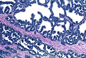
Background imageImages Dated 31st October 2007: Prostate gland, light micrograph

Prostate gland, light micrograph
Prostate gland. Coloured light micrograph of a section through a prostate gland. The prostate is a large male gland that surrounds the neck of the bladder and the urethra

Background imageImages Dated 31st October 2007: Oesophagus wall, light micrograph

Oesophagus wall, light micrograph
Oesophagus wall. Coloured light micrograph of a section through the human oesophagus, which passes food from the mouth to the stomach

Background imageImages Dated 31st October 2007: Stomach lining, light micrograph

Stomach lining, light micrograph
Stomach lining. Coloured light micrograph of the lining of the stomach, known as the mucosa. The stomach is a muscular sac involved in storage and digestion of food. The lumen is at top (white)

Background imageImages Dated 31st October 2007: Stomach lining, SEM

Stomach lining, SEM
Stomach lining. Coloured scanning electron micrograph (SEM) of the lining of the stomach, known as the mucosa. The stomach is a muscular sac involved in storage and digestion of food

Background imageImages Dated 31st October 2007: Oesophagus wall, light micrograph

Oesophagus wall, light micrograph
Oesophagus wall. Coloured light micrograph of a section through the human oesophagus, which passes food from the mouth to the stomach

Background imageImages Dated 31st October 2007: Nose mucosa, light micrograph

Nose mucosa, light micrograph
Nose mucosa. Light micrograph of a section through the nasal mucosa (the tissue that lines the airways of the nose), in the region lying over the nasal concha

Background imageImages Dated 31st October 2007: Lymph node, light micrograph

Lymph node, light micrograph
Lymph node. Coloured light micrograph of a section through a lymph node. Lymph nodes are kidney shaped encapsulated structures that are usually located at intervals along lymph vessels

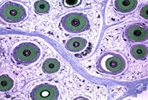
Background imageImages Dated 31st October 2007: Hair follicles, light micrograph

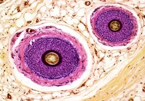
Hair follicles, light micrograph
Hair follicles. Coloured light micrograph of a section through hairs of the human skin. Two hair shafts (circular structures) are seen embedded in the dermis (white & brown) of the skin

Background imageImages Dated 31st October 2007: Ovarian follicle, light micrograph

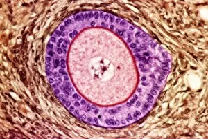
Ovarian follicle, light micrograph
Ovarian follicle. Coloured light micrograph of a section through a primary follicle in an ovary. At centre is the large nucleus of the developing egg (oocyte, pink)

Background imageImages Dated 31st October 2007: Ovarian follicles, light micrograph

Ovarian follicles, light micrograph
Ovarian follicles. Coloured light micrograph of a section through an ovary (orange), showing three primary follicles (pink)

Background imageImages Dated 31st October 2007: Internal wall of uterus, SEM

Internal wall of uterus, SEM
Internal wall of uterus. Coloured scanning electron micrograph of the mucosa, also known as endometrium, lining the body of the human uterus

Background imageImages Dated 31st October 2007: Salivary gland, light micrograph

Salivary gland, light micrograph
Salivary gland. Coloured light micrograph of a section through a sublingual salivary gland, which is situated directly under the tongue

Background imageImages Dated 31st October 2007: Salivary gland, light micrograph

Salivary gland, light micrograph
Salivary gland. Coloured light micrograph of a section through a sublingual salivary gland, which is situated directly under the tongue

Background imageImages Dated 31st October 2007: Salivary gland, light micrograph

Salivary gland, light micrograph
Salivary gland. Coloured light micrograph of a section through a sublingual salivary gland, which is situated directly under the tongue

Background imageImages Dated 31st October 2007: Salivary gland, light micrograph

Salivary gland, light micrograph
Salivary gland. Coloured light micrograph of a section through a sublingual salivary gland, which is situated directly under the tongue

Background imageImages Dated 31st October 2007: Salivary gland, light micrograph

Salivary gland, light micrograph
Salivary gland. Coloured light micrograph of a section through a submandibular salivary gland, one of a pair of salivary glands situated beneath the base of the tongue

Background imageImages Dated 31st October 2007: Mouse tongue, SEM

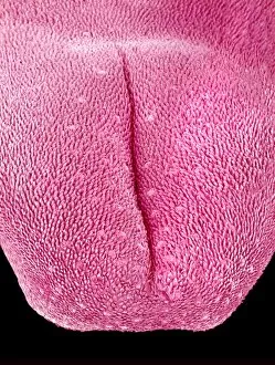
Mouse tongue, SEM
Tongue of a mouse, coloured scanning electron micrograph (SEM). The tongue is covered in thin projections called filiform papillae. These sense touch

Background imageImages Dated 31st October 2007: Tongue papillae, light micrograph

Tongue papillae, light micrograph
Tongue papillae. Coloured light micrograph of a section through the tongue, showing its papillae (brown). They project from the tongues upper surface, giving it its rough texture

Background imageImages Dated 31st October 2007: Tongue, light micrograph

Tongue, light micrograph
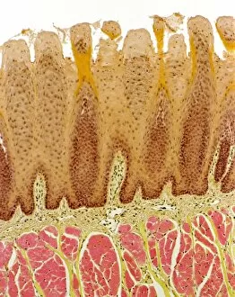
Tongue. Coloured light micrograph of a section through a tongue, showing its papillae (red). They project from the tongues upper surface, giving it its rough texture

Background imageImages Dated 31st October 2007: Tongue papillae, light micrograph

Tongue papillae, light micrograph
Tongue papillae. Coloured light micrograph of a section through the tongue, showing its filiform papillae (red). They project from the tongues upper surface, giving it its rough texture

Background imageImages Dated 31st October 2007: Taste bud, light micrograph

Taste bud, light micrograph
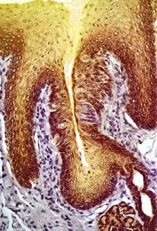
Taste bud. Coloured light micrograph of a section through the tongue, showing a taste bud. This taste bud is seen as a round brown mass slightly to the right of the vertical white line

Background imageImages Dated 31st October 2007: Taste bud, light micrograph

Taste bud, light micrograph
Taste bud. Coloured light micrograph of a section through the tongue, showing a taste bud. This taste bud is seen as a round purple mass slightly to the right of the vertical white line

Background imageImages Dated 31st October 2007: Retina, light micrograph

Retina, light micrograph
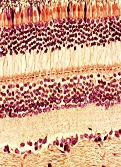
Retina. Coloured light micrograph of a section through a retina. The eye works by allowing light to be focused by the lens onto the retina

Background imageImages Dated 31st October 2007: Eye structure, light micrograph

Eye structure, light micrograph
Eye structure. Coloured light micrograph of a section through the inner surface of a human eye. This slice shows the vitreous humour (transparent liquid in the eye, top), the retina (pink)

Background imageImages Dated 31st October 2007: Nerve fibres, light micrograph

Nerve fibres, light micrograph
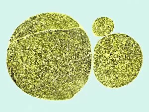
Nerve fibres. Coloured light micrograph of a transverse section through three bundles (fascicles) of nerve fibres. Within each fascicle are many myelinated nerve fibres

Background imageImages Dated 31st October 2007: Spine anatomy

Spine anatomy. Coloured light micrograph of a section through a spine, showing vertebra bone (purple), the spinal cord (yellow, upper centre), the spinal canal (white) and surrounding tissue (brown)

Background imageImages Dated 31st October 2007: Footprint recovery

Footprint recovery
MODEL RELEASED. Footprint recovery. Forensics officer photographing a footprint at crime scene. After taking the photograph, a cast will be made of the print

Background imageImages Dated 31st October 2007: Collecting evidence

Collecting evidence
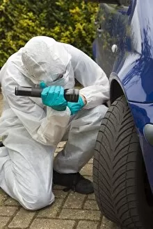
MODEL RELEASED. Collecting evidence. Scene of crime officer collecting evidence from a car tyre at a crime scene

Background imageImages Dated 31st October 2007: Forensic evidence

Forensic evidence
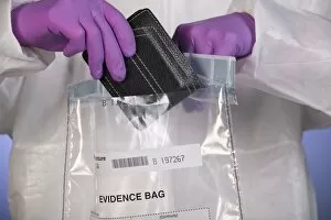
MODEL RELEASED. Forensic evidence. Forensic officer placing a wallet found at a crime scene in a police evidence bag for analysis

Background imageImages Dated 31st October 2007: Collecting evidence

Collecting evidence
MODEL RELEASED. Collecting evidence. Scene of crime officer collecting evidence from a car tyre at a crime scene

Background imageImages Dated 31st October 2007: Forensic evidence

Forensic evidence
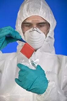
MODEL RELEASED. Forensic evidence. Forensic officer using a pair of tweezers to place a match box into an evidence bag

Background imageImages Dated 31st October 2007: Forensic evidence

Forensic evidence
MODEL RELEASED. Forensic evidence. Forensic officer placing a tool found at a crime scene in a police evidence bag for analysis

Background imageImages Dated 31st October 2007: Footprint recovery

Footprint recovery
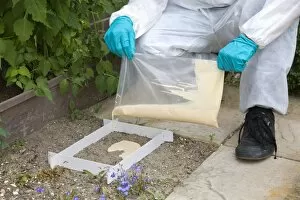
MODEL RELEASED. Footprint recovery. Forensics officer pouring fine plaster of paris over a footprint at a crime scene. This is done to create a permanent record of the print

Background imageImages Dated 31st October 2007: Fingerprint dusting

Fingerprint dusting
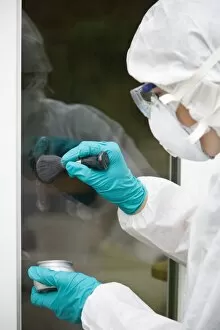
MODEL RELEASED. Fingerprint dusting. Forensics officer dusting a window for fingerprints at a crime scene

Background imageImages Dated 31st October 2007: Hair follicles, light micrograph

Hair follicles, light micrograph
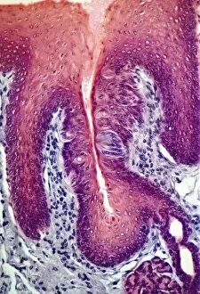
Hair follicles. Coloured light micrograph of a section through hairs of the human skin. Many hair shafts (circular structures) are seen embedded in the dermis (white) of the skin

Background imageImages Dated 31st October 2007: Buttercup stem, light micrograph

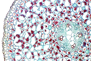
Buttercup stem, light micrograph
Buttercup stem. Coloured light micrograph of a section through a buttercup stem (Ranunculus sp.) showing the circular-shaped vascular bundle (right)

Background imageImages Dated 31st October 2007: Comfrey flower petal, SEM

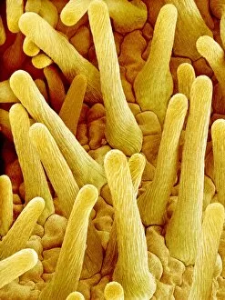
Comfrey flower petal, SEM
Comfrey flower petal. Coloured scanning electron micrograph (SEM) showing papillae on a petal from a comfrey (Symphytum officinale) flower. Papillae are projections from epidermal cells

Background imageImages Dated 31st October 2007: Morning glory pollen grains, SEM

Morning glory pollen grains, SEM
Morning glory pollen grains. Coloured scanning electron micrograph (SEM) of pollen grains from a morning glory flower (Ipomoea sp.)

Background imageImages Dated 31st October 2007: Silent Film Still: Sports. Guy Kibbee

Silent Film Still: Sports. Guy Kibbee
SILENT FILM STILL: SPORTS. Guy Kibbee

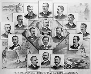
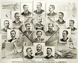
Background imageImages Dated 31st October 2007: BASEBALL, 1894. Representatives of professional baseball in America. Lithograph, 1894

BASEBALL, 1894. Representatives of professional baseball in America. Lithograph, 1894

Background imageImages Dated 31st October 2007: BICYCLE AD, 1896. English newspaper advertisment, 1896, for the Columbia bicycle

BICYCLE AD, 1896. English newspaper advertisment, 1896, for the Columbia bicycle, a brand imported from the United States

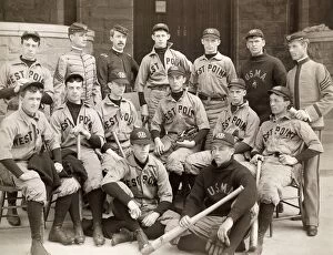
Background imageImages Dated 31st October 2007: BASEBALL: WEST POINT, 1896. The U. S. Military Academy baseball team, 1896, at West Point, New York

BASEBALL: WEST POINT, 1896. The U. S. Military Academy baseball team, 1896, at West Point, New York
BASEBALL: WEST POINT, 1896. The U.S. Military Academy baseball team, 1896, at West Point, New York

Background imageImages Dated 31st October 2007: BASEBALL, 1894. Representatives of professional baseball in America. Lithograph, 1894

BASEBALL, 1894. Representatives of professional baseball in America. Lithograph, 1894

Background imageImages Dated 31st October 2007: Ordesa Valley - Ordesa y Monte Perdido National Park - Pyrenees - Spain

Ordesa Valley - Ordesa y Monte Perdido National Park - Pyrenees - Spain
WAT-15878 Ordesa Valley - Ordesa y Monte Perdido National Park Pyrenees - Spain M. Watson Please note that prints are for personal display purposes only and may not be reproduced in any way

Background imageImages Dated 31st October 2007: Lammergeier / Bearded Vulture. Pyrenees - France / Europe

Lammergeier / Bearded Vulture. Pyrenees - France / Europe
WAT-15866 Lammergeier / Bearded Vulture. Pyrenees - France / Europe Gypaetus barbatus M. Watson Please note that prints are for personal display purposes only and may not be reproduced in any way


All Professionally Made to Order for Quick Shipping


-

Why Choose Us?

We are a leading provider of Art Prints and Photo Gifts since 2003, working in partnership with a range of Sporting Clubs, Charities, Museums and Picture Libraries. A large share of profits from any of their images will go directly towards supporting that charity or club. Our archive of images is carefully curated to bring you a wide range of subjects, including landscapes, wildlife, architecture, and more. We ship from our partner labs in the UK, USA, EU (Netherlands) and Australia.
+

How do I place an order?

Ordering is quick & easy - Just follow 5 Simple Steps:
  1. Find your image: Use our search box or browse our online photo Collections to find the image you want.

  2. Choose your print format: Select your desired product and add it to your cart.

  3. Enter your details: If you're a returning customer, simply enter your email address and password, and we'll fill in your billing and shipping address details. All personal details are held securely and are fully GDPR compliant. As standard, we remove all Personally Identifiable Information after 12 months.

  4. Pay for your purchase: We use state-of-the-art security for online shopping and do not have access to your card details.

  5. Sit back and relax: We'll email you confirmation of your order and when it's dispatched. Registered customers can also track orders in the 'My Account' area.

+

How do I pay for an order?

You can pay for your order with most of the major credit and debit cards, or PayPal. For added security, major financial institutions process payment details separately and securely on our behalf. We do not have access to your online payment card details. Online payments are preferred however we do take phone orders. For UK customers only we accept cheques issued against a UK bank.
+

Is my data safe?

We take data security very seriously. We do not have access to your full card details and all payments are requested over a fully secure connection. Additionally, we fully comply with current European and GDPR legislation, and automatically remove all personal data after at most 12 months (unless you have an account with us and have signed in recently). We also have a strict opt-in policy and would never sell your personal details. Your data is only used to fulfill your order promptly and efficiently.
+

Is wall art ready to hang?

For quick and easy installation all wall art, including framed prints, canvas prints and metal prints are supplied with a ready to hang solution on the back. Generally, saw tooth hangers are applied as they allow wall art to hang flush against the wall. The serrated edge of the hanger prevents the frame from shifting or tilting when hung.
+

Are the photo prints fade resistant?

Yes, we use archival quality photo paper photographic paper for vivid reproduction Prints are an accurate representations of the original artwork, which is preserved for artistic character and authenticity. We guarantee they match previews shown on our web site