Canvas Print : Chrysanthemum pistil, SEM


![]()
Canvas Prints from Science Photo Library
Chrysanthemum pistil, SEM
Chrysanthemum pistil. Coloured scanning electron micrograph (SEM) of a pistil from a Chrysanthemum sp. flower. The pistil is the female reproductive part of a plant. It comprises the stigma (yellow and orange, top), the style (orange, bottom) and the ovary (not seen). Pollen grains, a flowers male reproductive cells, land on the stigma and burrow a tube down to the ovary, where they fertilise the female sex cell (ovule). At bottom are the anthers, the pollen-producing part of the flowers male reproductive system. Magnification: x40 when printed at 10 centimetres wide
Science Photo Library features Science and Medical images including photos and illustrations
Media ID 6288533
© STEVE GSCHMEISSNER/SCIENCE PHOTO LIBRARY
Anther Carpel Disc Floret False Colour Fertile Pistil Re Production Reproductive Stigma Style False Coloured
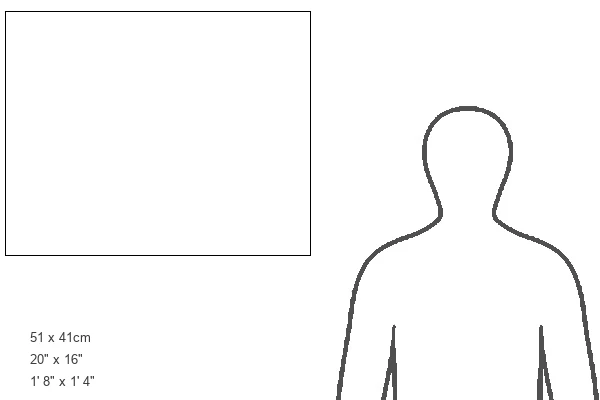
20"x16" (51x41cm) Canvas Print
Discover the intricacies of nature with our Media Storehouse Canvas Prints featuring "Chrysanthemum pistil, SEM" by Science Photo Library. This captivating image showcases the beauty and complexity of a Chrysanthemum pistil in stunning detail, captured through the advanced technology of Scanning Electron Microscopy. Each print is meticulously crafted on high-quality canvas, ensuring vibrant colors and exceptional clarity that bring the wonders of science right into your home or office. Impress your guests with this unique and thought-provoking art piece that combines art and science in perfect harmony.
Delivered stretched and ready to hang our premium quality canvas prints are made from a polyester/cotton blend canvas and stretched over a 1.25" (32mm) kiln dried knot free wood stretcher bar. Packaged in a plastic bag and secured to a cardboard insert for safe transit.
Canvas Prints add colour, depth and texture to any space. Professionally Stretched Canvas over a hidden Wooden Box Frame and Ready to Hang
Estimated Product Size is 50.8cm x 40.6cm (20" x 16")
These are individually made so all sizes are approximate
Artwork printed orientated as per the preview above, with landscape (horizontal) orientation to match the source image.
EDITORS COMMENTS
This print showcases the intricate beauty of a Chrysanthemum pistil, captured through a coloured scanning electron microscope (SEM). The pistil, which is the female reproductive part of the flower, is composed of three main components: the stigma, style, and ovary. In this image, we can observe the vibrant yellow and orange stigma at the top, followed by the orange-hued style at the bottom. Unfortunately, due to its microscopic nature, we cannot see the ovary in this particular shot. The fascinating process of plant reproduction unfolds within this tiny structure. Pollen grains from male flowers land on the stigma and penetrate down into a tube that leads to fertilization in the ovary. This fusion occurs when pollen reaches an egg cell (ovule), resulting in new life. At closer inspection towards the bottom of this image are anthers - responsible for producing pollen as part of a flower's male reproductive system. The magnification level used for printing was x40 when measured at 10 centimeters wide. Through this stunning photograph provided by Science Photo Library, we gain insight into both botanical beauty and scientific wonderment. It serves as a reminder that even within seemingly small details lies immense complexity and natural marvels waiting to be explored.
MADE IN AUSTRALIA
Safe Shipping with 30 Day Money Back Guarantee
FREE PERSONALISATION*
We are proud to offer a range of customisation features including Personalised Captions, Color Filters and Picture Zoom Tools
SECURE PAYMENTS
We happily accept a wide range of payment options so you can pay for the things you need in the way that is most convenient for you
* Options may vary by product and licensing agreement. Zoomed Pictures can be adjusted in the Cart.