Canvas Print : Heart valve tendons, SEM


![]()
Canvas Prints from Science Photo Library
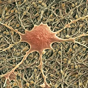
Heart valve tendons, SEM
Heart valve tendons. Coloured scanning electron micrograph (SEM) of the chordae tendineae, tendons which attach the atrioventricular heart valves to their muscles. These valves prevent blood being forced back into the atria (upper heart chambers) from the ventricles (lower heart chambers) during the contraction of the ventricles. The right ventricle (which pumps deoxygenated blood to the lungs) is separated from the right atrium by the tricuspid valve. The left ventricle (which pumps newly-oxygenated blood around the body) is separated from the left atrium by the mitral valve. The valves are controlled by papillary muscles (not seen). Magnification unknown
Science Photo Library features Science and Medical images including photos and illustrations
Media ID 6421249
© STEVE GSCHMEISSNER/SCIENCE PHOTO LIBRARY
Cardiac Chordae Tendineae Cord String Strings Tendon Tendons Tissue Valve Valves Cords
20"x16" (51x41cm) Canvas Print
Discover the intricacy of life with our Media Storehouse Canvas Prints featuring the captivating image "Heart valve tendons, SEM" by Science Photo Library. This mesmerizing scanning electron micrograph (SEM) reveals the intricate details of the chordae tendineae, the tendons that attach the atrioventricular heart valves to their muscles. Bring the beauty of science into your home or office with this high-quality canvas print, a stunning conversation starter that celebrates the wonders of the human body.
Delivered stretched and ready to hang our premium quality canvas prints are made from a polyester/cotton blend canvas and stretched over a 1.25" (32mm) kiln dried knot free wood stretcher bar. Packaged in a plastic bag and secured to a cardboard insert for safe transit.
Canvas Prints add colour, depth and texture to any space. Professionally Stretched Canvas over a hidden Wooden Box Frame and Ready to Hang
Estimated Product Size is 50.8cm x 40.6cm (20" x 16")
These are individually made so all sizes are approximate
Artwork printed orientated as per the preview above, with landscape (horizontal) orientation to match the source image.
EDITORS COMMENTS
This print showcases the intricate beauty of heart valve tendons, as seen through a colored scanning electron microscope (SEM). The chordae tendineae, represented here in stunning detail, are the tendons responsible for attaching the atrioventricular heart valves to their respective muscles. These valves play a crucial role in preventing blood from being forced back into the upper heart chambers during ventricular contractions. The image highlights two specific valves: the tricuspid valve separating the right ventricle from the right atrium and the mitral valve separating the left ventricle from the left atrium. While papillary muscles controlling these valves remain unseen, their importance cannot be understated. Through this SEM capture, we gain insight into both healthy anatomy and cardiac function. The magnification level remains unknown but serves to emphasize just how remarkable these tiny structures are within our bodies. The vibrant colors bring attention to every string-like tendon and cord that forms this complex network within our hearts. It is a testament to nature's design and reminds us of its incredible intricacy at even microscopic levels. This print by Science Photo Library offers an awe-inspiring glimpse into human biology while showcasing both scientific curiosity and artistic appreciation for our own inner workings.
MADE IN AUSTRALIA
Safe Shipping with 30 Day Money Back Guarantee
FREE PERSONALISATION*
We are proud to offer a range of customisation features including Personalised Captions, Color Filters and Picture Zoom Tools
SECURE PAYMENTS
We happily accept a wide range of payment options so you can pay for the things you need in the way that is most convenient for you
* Options may vary by product and licensing agreement. Zoomed Pictures can be adjusted in the Cart.