Fraud Blocker Skip to main content

Abdominal Collection

Background imageAbdominal Collection: Athletic young man in shorts with ball lying on a stone floor

Athletic young man in shorts with ball lying on a stone floor

Background imageAbdominal Collection: Cardiovascular system, historical artwork

Cardiovascular system, historical artwork. 19th Century hand coloured lithographic print showing the blood vessels (red and blue) of the human neck and torso

Background imageAbdominal Collection: Young man in a suit and without shirt lying on a couch

Young man in a suit and without shirt lying on a couch

Background imageAbdominal Collection: Young man in a suit and without shirt kneeling on a couch

Young man in a suit and without shirt kneeling on a couch

Background imageAbdominal Collection: Man with a naked torso wearing long underwear in a horse barn sitting on bales of hay

Man with a naked torso wearing long underwear in a horse barn sitting on bales of hay

Background imageAbdominal Collection: Male groin arteries, 1825 artwork

Male groin arteries, 1825 artwork
Male groin arteries. Dissections of a male groin to show areas supplied by the internal iliac artery (red) and its branches

Background imageAbdominal Collection: Abdominal organs

Abdominal organs, historical anatomical artwork. Several of the organs involved in digestion have been removed from the abdomen. The liver (brown) is at upper left, and a kidney (red) at lower right

Background imageAbdominal Collection: Woman using an exercise ball

Woman using an exercise ball
MODEL RELEASED. Woman using an exercise ball (Swiss ball) to do sit-ups. A variety of exercises can be performed with this piece of equipment. It is used in the Pilates exercise system

Background imageAbdominal Collection: Cute african american girl wearing casual clothes with hand on stomach because indigestion

Cute african american girl wearing casual clothes with hand on stomach because indigestion, painful illness feeling unwell. ache concept.

Background imageAbdominal Collection: Internal organs of the abdomen and thorax (colour litho)

Internal organs of the abdomen and thorax (colour litho)
3097100 Internal organs of the abdomen and thorax (colour litho) by German School, (19th century); Private Collection; (add.info.: Internal organs of the abdomen and thorax)

Background imageAbdominal Collection: Upset woman and her narcissistic boyfriend Upset woman and her narcissistic boyfriend

Upset woman and her narcissistic boyfriend Upset woman and her narcissistic boyfriend

Background imageAbdominal Collection: Young Man Standing Strong In The Gym And Flexing Muscles - Muscular Athletic Bodybuilder Fitness Model Posing After

Young Man Standing Strong In The Gym And Flexing Muscles - Muscular Athletic Bodybuilder Fitness Model Posing After Exercises

Background imageAbdominal Collection: Human digestive system, digestive tract or alimentary canal including labels with US spellings

Human digestive system, digestive tract or alimentary canal including labels with US spellings

Background imageAbdominal Collection: Doctor doing a thoracic and abdominal recognition of a patie

Doctor doing a thoracic and abdominal recognition of a patie
History of medicine. Doctor doing a thoracic and abdominal recognition of a patient. Miniature. Anatomy treaty. 14th century

Background imageAbdominal Collection: Naked man holding a blank sign in his hands

Naked man holding a blank sign in his hands

Background imageAbdominal Collection: Young athlete in white briefs

Young athlete in white briefs

Background imageAbdominal Collection: Lawson Tait

Lawson Tait
3586304 Lawson Tait; (add.info.: Photograph of Lawson Tait (1845-1899) a Scottish pioneer in pelvic and abdominal surgery. Dated 19th Century.); Universal History Archive/UIG

Background imageAbdominal Collection: The abdominal aorta and its branches (engraving)

The abdominal aorta and its branches (engraving)
3634189 The abdominal aorta and its branches (engraving) by English School, (19th century); Private Collection; (add.info.: The abdominal aorta and its branches)

Background imageAbdominal Collection: Bandages, wrappings and compresses (colour litho)

Bandages, wrappings and compresses (colour litho)
3097096 Bandages, wrappings and compresses (colour litho) by German School, (19th century); Private Collection; (add.info.: Bandages, wrappings and compresses)

Background imageAbdominal Collection: Anatomy, the abdominal viscera

Anatomy, the abdominal viscera
5202581 Anatomy, the abdominal viscera, kidneys, etc, the oestrus equi, gadfly, ova, larvae, bots etc (litho) by Herring, Benjamin (1830-71); Private Collection; (add.info.: Anatomy)

Background imageAbdominal Collection: Topography of thoracic and abdominal viscera (engraving)

Topography of thoracic and abdominal viscera (engraving)
3634177 Topography of thoracic and abdominal viscera (engraving) by English School, (19th century); Private Collection; (add.info.: Topography of thoracic and abdominal viscera)

Background imageAbdominal Collection: Vintage Anatomical Print of the Human Thoracic and Abdominal Regions, 1912 (screen print)

Vintage Anatomical Print of the Human Thoracic and Abdominal Regions, 1912 (screen print)
3034949 Vintage Anatomical Print of the Human Thoracic and Abdominal Regions, 1912 (screen print) by American School

Background imageAbdominal Collection: Page from Surgical instrument catalogue, c. 1900: Abdominal belts and trusses (litho)

Page from Surgical instrument catalogue, c. 1900: Abdominal belts and trusses (litho)
6033268 Page from Surgical instrument catalogue, c.1900: Abdominal belts and trusses (litho) by English School, (20th century); Private Collection; (add.info.: Abdominal belts and trusses)

Background imageAbdominal Collection: Normal situation of the internal organs of the abdomen in men and women (engraving)

Normal situation of the internal organs of the abdomen in men and women (engraving)
3097160 Normal situation of the internal organs of the abdomen in men and women (engraving) by German School

Background imageAbdominal Collection: Bare-chested man in jeans

Bare-chested man in jeans

Background imageAbdominal Collection: Bare-chested man in jeans, kneeling

Bare-chested man in jeans, kneeling

Background imageAbdominal Collection: Athletic man, chest, front, chest, abdomen

Athletic man, chest, front, chest, abdomen

Background imageAbdominal Collection: Man with a naked torso wearing a pair of jeans

Man with a naked torso wearing a pair of jeans

Background imageAbdominal Collection: Masculine man without a shirt, wearing a cowboy hat and holding a guitar on a beach

Masculine man without a shirt, wearing a cowboy hat and holding a guitar on a beach

Background imageAbdominal Collection: Ovarian cyst, endoscope view C017 / 6800

Ovarian cyst, endoscope view C017 / 6800
Ovarian cyst. Endoscope (laparascope) view of a cyst (white) on a patients ovary. Ovarian cysts are common in women of child-bearing age

Background imageAbdominal Collection: Uterus, endoscope view C017 / 6805

Uterus, endoscope view C017 / 6805
Uterus. Endoscope (laparascope) view of a healthy uterus (womb, large, upper centre), with the two fallopian tubes (left and right) and an ovary (white, lower right)

Background imageAbdominal Collection: Female skeleton and organs, artwork

Female skeleton and organs, artwork
Female skeleton and organs and blood vessels, artwork

Background imageAbdominal Collection: Female anatomy, historical model

Female anatomy, historical model
Female anatomy. 1920s anatomical model showing the internal organs of the female upper body with the intestines, stomach and liver removed

Background imageAbdominal Collection: Male torso skeleton

Male torso skeleton. The bones of the chest and abdomen surround and protect the organs and provide support for the body. The ribcage (upper frame) encloses the heart and lungs

Background imageAbdominal Collection: Female abdominal anatomy, artwork

Female abdominal anatomy, artwork
Female abdominal anatomy. Computer artwork showing the kidneys and female reproductive system. Part of the lymphatic system (green) is also shown

Background imageAbdominal Collection: Comic German postcard -- health spa new invention

Comic German postcard -- health spa new invention
Comic German postcard by Arthur Thiele -- greetings from Karlsbad -- a grossly overweight man uses a new invention for abdominal support, looking very much like a wheelbarrow. Date: early 20th century

Background imageAbdominal Collection: Mittens and body belt, WW1 knitting, comforts for troops

Mittens and body belt, WW1 knitting, comforts for troops
Patterns for mittens and a body belt (designed to keep the midriff warm)

Background imageAbdominal Collection: Young athlete in jeans, with half-naked chest

Young athlete in jeans, with half-naked chest

Background imageAbdominal Collection: Muscular man wearing swimming trunks standing on a beach

Muscular man wearing swimming trunks standing on a beach

Background imageAbdominal Collection: Diaphragm anatomy, 1831 artwork

Diaphragm anatomy, 1831 artwork
Diaphragm anatomy, posterior view. The diaphragm is the white area at top, connected to surrounding muscles (red). Part of the spine is at bottom, and ribs at left and right

Background imageAbdominal Collection: Ovary, endoscope view C017 / 6803

Ovary, endoscope view C017 / 6803
Ovary. Endoscope (laparascope) view of a healthy ovary (white). There are two ovaries in the female body, which release an egg (ovum) during the monthly ovulation cycle

Background imageAbdominal Collection: Abdominal organs, 1880 artwork C017 / 6912

Abdominal organs, 1880 artwork C017 / 6912
Abdominal organs. Historical anatomical artwork of a dissected abdomen seen from the front, showing the internal organs. These include the intestines, bladder, stomach and liver

Background imageAbdominal Collection: Trunk muscle anatomy, 1831 artwork

Trunk muscle anatomy, 1831 artwork
Trunk muscle anatomy. Coronal section of the trunk (anterior view) showing the anatomy of its muscles and bones

Background imageAbdominal Collection: Male skeleton and organs, artwork

Male skeleton and organs, artwork
Male skeleton and organs and blood vessels, artwork

Background imageAbdominal Collection: Vagus nerve anatomy, artwork C018 / 0300

Vagus nerve anatomy, artwork C018 / 0300
Vagus nerve anatomy. Artwork of a human body from the side, showing the internal anatomy and the location of the vagus nerve (yellow)

Background imageAbdominal Collection: Ant anus, SEM C018 / 0550

Ant anus, SEM C018 / 0550
Ant anus, coloured scanning electron micrograph (SEM). Ants excrete bodily waste through a single orifice, their anus, which is located at the tip of the abdomen

Background imageAbdominal Collection: Female groin arteries, 1825 artwork

Female groin arteries, 1825 artwork
Female groin arteries. Dissections of a female groin to show areas supplied by the internal iliac artery (red) and its branches

Background imageAbdominal Collection: Trunk musculature, 1831 artwork

Trunk musculature, 1831 artwork
Trunk musculature, anterior view. This anatomical artwork is plate 62 from volume 2 (1831) of Traite complet de l anatomie de l homme (1831-1854)

Background imageAbdominal Collection: Human anatomy, 1823 C017 / 8057

Human anatomy, 1823 C017 / 8057
Human anatomy. 19th-century illustrations showing the anatomy of a human skull (upper left), brain and spinal column (upper centre), chest and abdominal organs (lungs, heart, liver, stomach)

Background imageAbdominal Collection: Twisted colon, X-ray C017 / 7752

Twisted colon, X-ray C017 / 7752
Twisted colon. X-ray of the abdomen of a 68 year old male patient with a twisted sigmoid colon, a condition known as a volvulus

Background imageAbdominal Collection: False-colour CT scan of liver & stomach

False-colour CT scan of liver & stomach
False-colour computed X-ray tomography (CT) scan of an axial section through the upper abdomen showing a normal liver (left, mostly yellow) and stomach (right, blue, with black part at top)

Background imageAbdominal Collection: Patient examination, 14th century artwork

Patient examination, 14th century artwork
Patient examination. 14th century artwork of a doctor examining a patient by palpation of the abdomen. Artwork from Anathomia (1345), by the Italian anatomist Guido da Vigevano

Background imageAbdominal Collection: Ectopic hydrocephalic shunt, 3D MRI scan

Ectopic hydrocephalic shunt, 3D MRI scan
Ectopic hydrocephalic shunt. Coloured 3D magnetic resonance imaging (MRI) scan of the pelvis of a patient, showing an ectopic (abnormally positioned) hydrocephalic shunt (ring, centre)

Background imageAbdominal Collection: Female internal organs, artwork

Female internal organs, artwork
Lymphatic system. Computer artwork of a naked woman with a healthy lymphatic system. Also seen are associated organs including the thymus gland (upper centre) and the spleen (centre right)

Background imageAbdominal Collection: Pelvic pain, conceptual artwork

Pelvic pain, conceptual artwork
Pelvic pain. Conceptual computer artwork showing pain (represented as the red area) in pelvis

Background imageAbdominal Collection: Intefs seated statue. Egypt

Intefs seated statue. Egypt
Intefs seated statue. Limestone. Wearing with a wig, long skirt and with abdominal folds with which scuptor shows Intefs high social status, associated to abundant food and sedentary lifestyle

Background imageAbdominal Collection: WW1 knitting, woollen belts - gift to the troops from Queen

WW1 knitting, woollen belts - gift to the troops from Queen
Gift to the Troops at the Front from the Queen and the Women of the Empire. Leaflet giving instructions on how to knit woollen belts for troops during the First World War

Background imageAbdominal Collection: Sir Astley Paston Cooper

Sir Astley Paston Cooper (1768 - 1841), 1st Baronet, English surgeon and anatomist, surgeon to Guys Hospital. In 1820 he removed an infected sebaceous cyst from the head of King George IV

Background imageAbdominal Collection: Typhus Sufferers in Kniagin, Russia

Typhus Sufferers in Kniagin, Russia
Bedridden Typhus sufferers in the Russian provence of Kniagin. Date: 1891 - 1892

Background imageAbdominal Collection: Train Transporting Typhoid Sufferers

Train Transporting Typhoid Sufferers
A Russian train being used as a temporary hospital specialising in treatment for patients suffering from Typhoid fever. Date: 1920

Background imageAbdominal Collection: Frontal trunk anatomy engraving 1866

Frontal trunk anatomy engraving 1866
Atlas d anatomie descriptive du corps humain C. Bonamy - Paul Broca Victor Masson et Fils Paris 1866

Background imageAbdominal Collection: Sternum anatomy engraving 1866

Sternum anatomy engraving 1866
Atlas d anatomie descriptive du corps humain C. Bonamy - Paul Broca Victor Masson et Fils Paris 1866

Background imageAbdominal Collection: Bare-chested young man wearing pyjama bottoms lying in bed

Bare-chested young man wearing pyjama bottoms lying in bed

Background imageAbdominal Collection: Female blue-tailed damselfly (Ischnura elegans)

Female blue-tailed damselfly (Ischnura elegans), violet form, resting on a dried grass stem, Creech Heath, Dorset, England, United Kingdom, Europe

Background imageAbdominal Collection: Male blue-tailed damselfly (Ischnura elegans) resting on a bracken frond, Studland Heath

Male blue-tailed damselfly (Ischnura elegans) resting on a bracken frond, Studland Heath, Dorset, England, United Kingdom, Europe

Background imageAbdominal Collection: Hoverfly (Volucella bombylans var. plumata) visiting a greater knapweed flower (Centaurea

Hoverfly (Volucella bombylans var. plumata) visiting a greater knapweed flower (Centaurea scabiosa) in a chalk grassland meadow, Wiltshire, England, United Kingdom, Europe

Background imageAbdominal Collection: Male small red damselfly (Ceriagrion tenellum) infested with mites perched on a sedge stem

Male small red damselfly (Ceriagrion tenellum) infested with mites perched on a sedge stem, Creech Heath, Dorset, England, United Kingdom, Europe

Background imageAbdominal Collection: Abdominal cavity and its contents. From Andreas Vesalius De humani corporis fabrica ja

Abdominal cavity and its contents. From Andreas Vesalius De humani corporis fabrica ja, Basle, 1543. Engraving

Background imageAbdominal Collection: Theodor Billroth (1829-94) Austrian surgeon who practiced in Vienna

Theodor Billroth (1829-94) Austrian surgeon who practiced in Vienna. Pioneer of abdominal surgery: resection of intestine (1881), excision of larynx (1874)

Background imageAbdominal Collection: Illustration of mesentery, small intestine and colon

Illustration of mesentery, small intestine and colon
Medicine: Human anatomy, mesentery, small intestine and colon. Drawing

Background imageAbdominal Collection: Illustration of stomach, abdominal cavity

Illustration of stomach, abdominal cavity
Medicine: Human anatomy, stomach, abdominal cavity, position

Background imageAbdominal Collection: Illustration of abdominal organs in chest

Illustration of abdominal organs in chest
Medicine: Human anatomy, abdominal organs in the chest, topography

Background imageAbdominal Collection: Ovarian cyst, endoscope view C017 / 6801

Ovarian cyst, endoscope view C017 / 6801
Ovarian cyst. Endoscope (laparascope) view of a cyst (white) on a patients ovary. Ovarian cysts are common in women of child-bearing age

Background imageAbdominal Collection: Abdominal pain, artwork F007 / 7236

Abdominal pain, artwork F007 / 7236
Abdominal pain, computer artwork

Background imageAbdominal Collection: Female anatomy, artwork F008 / 3290

Female anatomy, artwork F008 / 3290
Female anatomy, computer artwork

Background imageAbdominal Collection: Female anatomy, artwork F008 / 3291

Female anatomy, artwork F008 / 3291
Female anatomy, computer artwork

Background imageAbdominal Collection: Human anatomy, artwork F008 / 3211

Human anatomy, artwork F008 / 3211
Human anatomy, artwork

Background imageAbdominal Collection: Human intestines, artwork

Human intestines, artwork
Human intestines. Artwork of a human torso with the intestines highlighted in colour. They consist of the small intestine (pink), circled by the large intestine (light brown)

Background imageAbdominal Collection: Intestinal veins, 1825 artwork

Intestinal veins, 1825 artwork
Intestinal veins. Dissection of the abdominal cavity showing the inferior mesenteric vein (blue) that drains blood from the large intestine

Background imageAbdominal Collection: Liver and stomach nerves, 1825 artwork

Liver and stomach nerves, 1825 artwork
Liver and stomach nerves. Extensive network of nerves (white) supplying the liver (upper left) and stomach (lower right)

Background imageAbdominal Collection: Intestinal arteries, 1825 artwork

Intestinal arteries, 1825 artwork
Intestinal arteries

Background imageAbdominal Collection: Abdominal aorta, 1825 artwork

Abdominal aorta, 1825 artwork
Abdominal aorta. Dissection, with some organs removed, showing the abdominal portion of the aorta (red), the main body artery

Background imageAbdominal Collection: Ovarian cyst, endoscope view C017 / 6802

Ovarian cyst, endoscope view C017 / 6802
Ovarian cyst. Endoscope (laparascope) view of a cyst (white) on a patients ovary. Ovarian cysts are common in women of child-bearing age

Background imageAbdominal Collection: Ectopic pregnancy, endoscope view C017 / 6804

Ectopic pregnancy, endoscope view C017 / 6804
Ectopic pregnancy. Endoscope (laparascope) view of an ectopic pregnancy in a patients fallopian tube


All products are expertly crafted, using premium materials, tailored to your specifications and promptly shipped


EDITORS COMMENTS

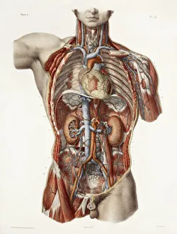
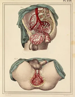
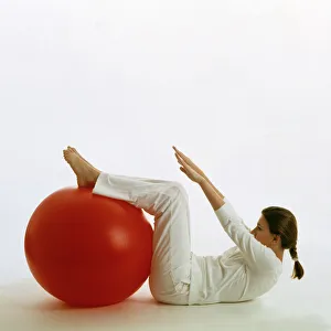
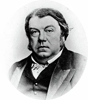
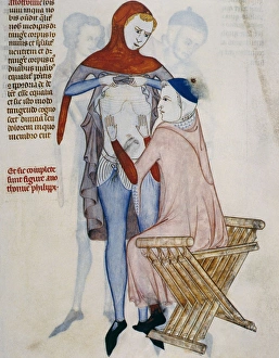
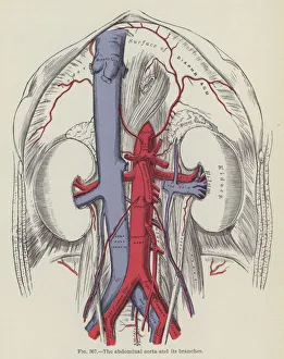
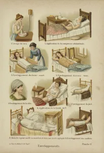
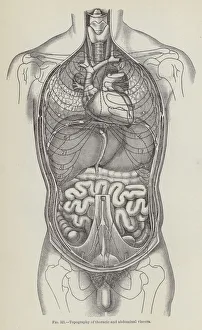
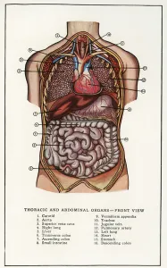
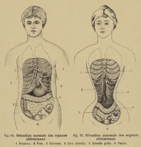
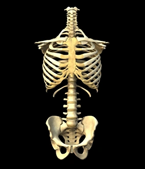
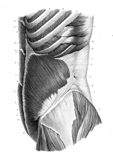
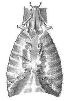
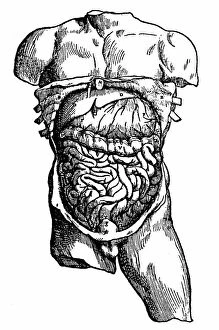
"Unleash the Power of Your Abdominal Strength: From Ancient Artwork to Modern Fitness" Athletic young man in shorts with a ball lying on a stone floor: This dedicated athlete knows that strong abdominal muscles are key to his performance and agility. Cardiovascular system meets historical artwork: Explore how the ancient Greeks depicted the importance of a well-toned abdomen in their sculptures, recognizing its connection to overall health and vitality. Young man in a suit and without shirt lying on a couch: Even amidst busy schedules, this gentleman understands the significance of maintaining core strength for good posture and stability. Young man in a suit and without shirt kneeling on a couch: Witness the dedication as this individual engages his abdominal muscles while working from home, demonstrating that fitness can be integrated into any routine. Male groin arteries, 1825 artwork: Delve into medical illustrations from centuries ago, showcasing early anatomical studies highlighting the intricate network of blood vessels within our abdominal region. Man with naked torso wearing long underwear in horse barn sitting on bales of hay: Embrace unconventional workout spaces as we witness this rugged individual utilizing his surroundings to engage his core muscles effectively. Woman using an exercise ball: Discover how incorporating an exercise ball into your fitness routine can target your abs while improving balance and flexibility for optimal results. Healthy lower back X-ray reveals strong abdominals' impact: Peek inside at an X-ray image showcasing how well-developed abdominal muscles provide support for our spine, preventing lower back pain or injuries. Abdominal organs - The hidden heroes within us all. Gain insight into these vital organs residing within our abdomen that play crucial roles in digestion, metabolism, immunity, and more. Sir Astley Paston Cooper's legacy lives on through understanding abdominals' importance - Learn about Sir Astley Paston Cooper's pioneering work in surgical anatomy, including his contributions to understanding the abdominal region.

© Copyright Media Storehouse, All Rights Reserved 2003 - 2025
Facebook     Pinterest     100% Payment Secure     Reviews IO