
Alveoli Collection
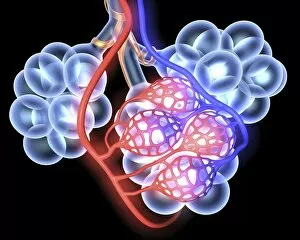
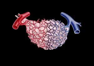
Alveoli, the tiny air sacs in our lungs, play a vital role in respiration


All Professionally Made to Order for Quick Shipping
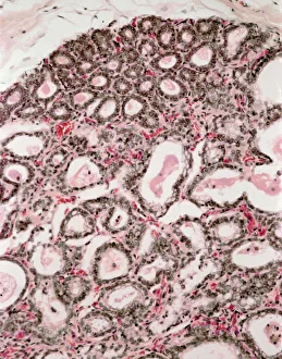
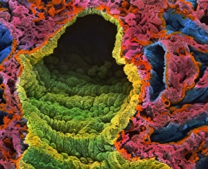
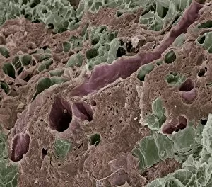
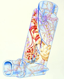
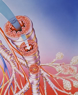
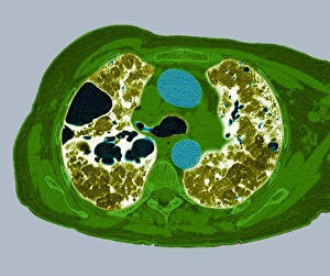
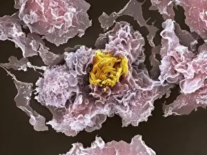
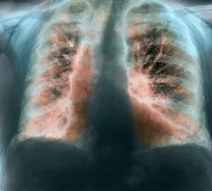
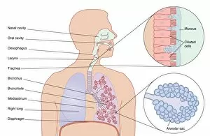
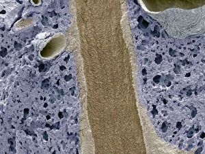
Alveoli, the tiny air sacs in our lungs, play a vital role in respiration. These grape-like structures are responsible for the exchange of oxygen and carbon dioxide between the bloodstream and the air we breathe. In healthy human lungs, alveoli form a complex network that allows efficient gas exchange. When viewed under a light micrograph, lactating breast tissue exhibits an intriguing resemblance to lung alveoli. This similarity highlights nature's remarkable ability to create specialized structures for different functions. However, not all it can healthy. In diseased lungs, such as those affected by bacterial infections or other respiratory conditions, these delicate sacs can become inflamed and damaged. The resulting impairment in their function can lead to breathing difficulties and reduced oxygen supply to the body. Artwork depicting lung alveoli anatomy showcases their intricate structure within our respiratory system. It emphasizes how these small but mighty units contribute to our overall well-being by ensuring proper gas exchange during each breath we take. Scanning electron microscopy (SEM) images provide detailed views of various aspects related to alveoli. From SEM images of lung alveoli themselves to blood vessels within the lung tissue, these visuals offer insights into their microscopic features and interactions with surrounding components. Understanding the importance of maintaining healthy alveolar function is crucial for preventing respiratory complications and promoting overall lung health.