Fraud Blocker Skip to main content

Anther Collection (page 4)

"Exploring the Intricate World of Anthers: A Captivating Microscopic Journey" Delve into the mesmerizing realm of anthers

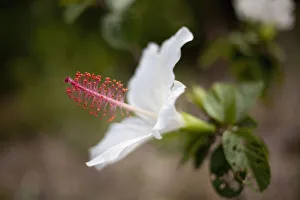
Background imageAnther Collection: Hawaii, Maui, A closeup of white hibiscus flower

Hawaii, Maui, A closeup of white hibiscus flower

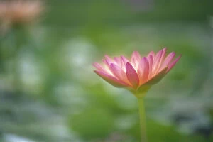
Background imageAnther Collection: Close-Up Side View Pink Lily In Pond, Green Blurry Background

Close-Up Side View Pink Lily In Pond, Green Blurry Background

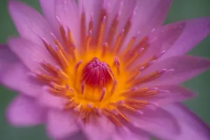
Background imageAnther Collection: Hawaii, Maui, Extreme Close-Up Pink Lily Blossom With Yellow Center, Detail

Hawaii, Maui, Extreme Close-Up Pink Lily Blossom With Yellow Center, Detail

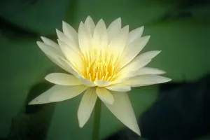
Background imageAnther Collection: Close-Up Of One White Lily Blossom In Pond, Green Leaves Background

Close-Up Of One White Lily Blossom In Pond, Green Leaves Background

Background imageAnther Collection: Light Micrograph (LM): Transverse section of Dehiscence Lilium Anthers with Pollen; Magnification

Light Micrograph (LM): Transverse section of Dehiscence Lilium Anthers with Pollen; Magnification x300 (on 10)
LRDS-200 Light Micrograph (LM): Transverse section of Dehiscence Lilium Anthers with Pollen Magnification x300 (on 10.5 cm width print) David Spears (Last Refuge)

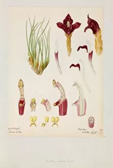
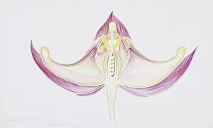
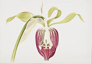
Background imageAnther Collection: Maxillaria subulata

Maxillaria subulata - By Franz Bauer, 1818. Paper, watercolour. Botany Library, Bauer Unit. Unfinished Drawings Monocotyledons 2, Orchidaceae 2, f. 149. Date: 1818
Background imageAnther Collection: Life cycle of a flowering plant

Life cycle of a flowering plant

Background imageAnther Collection: Ms Fr. Fv VI #1 fol. 149r Illustration from the Book of Simple Medicines

Ms Fr. Fv VI #1 fol. 149r Illustration from the Book of Simple Medicines
XJL212064 Ms Fr. Fv VI #1 fol.149r Illustration from the Book of Simple Medicines by Mattheaus Platearius (d.c.1161) c.1470 (vellum) by Testard, Robinet (fl.1470-1523); 26x35.5 cm; National Library

Background imageAnther Collection: Hybrid daylily stamen, consisting of anthers and filaments, Louisville, Kentucky

Hybrid daylily stamen, consisting of anthers and filaments, Louisville, Kentucky
Hybrid daylily stamen, consisting of anthers and, filaments, Louisville, Kentucky

Background imageAnther Collection: Picture No. 11014593

Picture No. 11014593
Peruvian Lily / Alstroemeria Pollen on Anther Scanning Electron Micrograph (SEM): Magnification x6520 (if print A4 size: 29.7 cm wide)

Background imageAnther Collection: Picture No. 10885015

Picture No. 10885015
Brown bee orchid - close-up - paticular of anther (Ophrys fusca) Date:

Background imageAnther Collection: Person removing anthers from yellow tomato flower, close-up

Person removing anthers from yellow tomato flower, close-up

Background imageAnther Collection: Hybridising roses, hands brushing anthers of pollinator across stigmas of seed parent, close-up

Hybridising roses, hands brushing anthers of pollinator across stigmas of seed parent, close-up

Background imageAnther Collection: Womans hands peeling off petals from rose, close-up

Womans hands peeling off petals from rose, close-up

Background imageAnther Collection: Hybridizing rose showing exposed anthers ready to release tiny pollen grains

Hybridizing rose showing exposed anthers ready to release tiny pollen grains

Background imageAnther Collection: Rubbing anthers of pollen parent over stigma of seed-bearing parent (cross-pollinating a tomato)

Rubbing anthers of pollen parent over stigma of seed-bearing parent (cross-pollinating a tomato), close-up

Background imageAnther Collection: Salix udensis Sekka (Willow) catkins and anthers on stem, close-up

Salix udensis Sekka (Willow) catkins and anthers on stem, close-up

Background imageAnther Collection: Spiderwort anther and pollen, SEM C017 / 1911

Spiderwort anther and pollen, SEM C017 / 1911
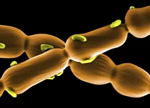
Spiderwort anther and pollen. Coloured scanning electron micrograph (SEM) of pollen grains (yellow) on hairs from the anther of a Spiderwort (Tradescantia sp.) flower

Background imageAnther Collection: Flowers reproductive structures, artwork

Flowers reproductive structures, artwork
Flowers reproductive structures, computer artwork. At centre is the female part of the flower, the carpel (green), which consists of a stigma (top), style (down centre) and ovary (bottom)

Background imageAnther Collection: Camellia flower stamens, SEM C015 / 9084

Camellia flower stamens, SEM C015 / 9084
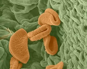
Camellia flower stamens. Coloured scanning electron micrograph (SEM) of the stamens of a Camellia sp. flower. Stamens are the male reproductive structures of a flower

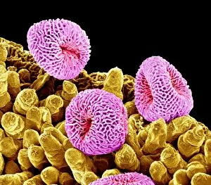
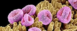
Background imageAnther Collection: Geranium pollen, SEM C015 / 9155

Geranium pollen, SEM C015 / 9155
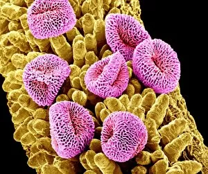
Geranium pollen. Coloured scanning electron micrograph (SEM) of pollen (pink) on the stamen of a geranium flower (Geranium sp.)

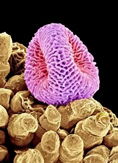
Background imageAnther Collection: Geranium pollen, SEM C015 / 9159

Geranium pollen, SEM C015 / 9159
Geranium pollen. Coloured scanning electron micrograph (SEM) of a pollen grain (pink) on the stamen of a geranium flower (Geranium sp.)

Background imageAnther Collection: Camellia flower stamens, SEM C015 / 9066

Camellia flower stamens, SEM C015 / 9066
Camellia flower stamens. Coloured scanning electron micrograph (SEM) of the stamens of a Camellia sp. flower. Stamens are the male reproductive structures of a flower

Background imageAnther Collection: Geranium pollen, SEM C015 / 9156

Geranium pollen, SEM C015 / 9156
Geranium pollen. Coloured scanning electron micrograph (SEM) of pollen (pink) on the stamen of a geranium flower (Geranium sp.)

Background imageAnther Collection: Geranium pollen, SEM C015 / 9157

Geranium pollen, SEM C015 / 9157
Geranium pollen. Coloured scanning electron micrograph (SEM) of pollen (pink) on the stamen of a geranium flower (Geranium sp.)

Background imageAnther Collection: Judas tree flower, SEM C016 / 0061

Judas tree flower, SEM C016 / 0061
Judas tree flower. Coloured scanning electron micrograph (SEM) of the reproductive parts of the flower of the Judas tree, or redbud (Cercis sp.)

Background imageAnther Collection: Geranium pollen, SEM C015 / 9158

Geranium pollen, SEM C015 / 9158
Geranium pollen. Coloured scanning electron micrograph (SEM) of a pollen grain (pink) on the stamen of a geranium flower (Geranium sp.)

Background imageAnther Collection: Judas tree flower, SEM C016 / 0062

Judas tree flower, SEM C016 / 0062
Judas tree flower. Coloured scanning electron micrograph (SEM) of the reproductive parts of the flower of the Judas tree, or redbud (Cercis sp.)

Background imageAnther Collection: Bee pollinating a flower, artwork C018 / 0297

Bee pollinating a flower, artwork C018 / 0297
Bee pollinating a flower. Sequence of artworks showing how a bee (left) lands on a flower to drink nectar (centre left), brushing up against the pollen-bearing anthers

Background imageAnther Collection: Flower structure, artwork C017 / 7232

Flower structure, artwork C017 / 7232
Flower structure. Computer artwork showing the main structures of a typical flower. The flower is the reproductive unit of some angiosperm plants

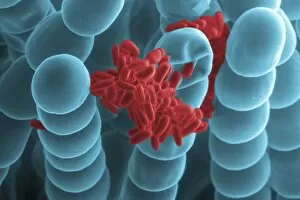
Background imageAnther Collection: Spiderwort anther and pollen, SEM C017 / 1910

Spiderwort anther and pollen, SEM C017 / 1910
Spiderwort anther and pollen. Coloured scanning electron micrograph (SEM) of hairs (blue) and pollen grains (red) on the anther of a Spiderwort (Tradescantia sp.) flower

Background imageAnther Collection: Reproductive organs of a lily flower

Reproductive organs of a lily flower
Flower of the hybrid Tango lily Cappuccino showing the reproductive organs. Lilies are monocotyledonous plants with three-fold symmetry in the floral parts.The picture shows 6 anthers (orange)

Background imageAnther Collection: Tulipa sp. flower, light micrograph C014 / 4654

Tulipa sp. flower, light micrograph C014 / 4654
Tulip (Tulipa sp.) flower. Light micrograph of a tulip flower, showing the male anthers (dark) and female stigma (centre). Magnification: x17 when printed 10 centimetres wide

Background imageAnther Collection: Crocus flower C014 / 4280

Crocus flower C014 / 4280
Crocus flower. Close-up of the stigma of a Crocus sp. flower

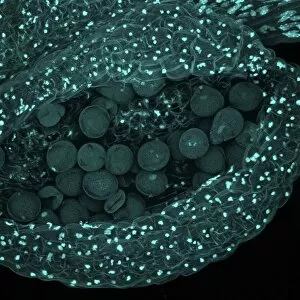
Background imageAnther Collection: Thale cress anther and pollen

Thale cress anther and pollen. Confocal laser scanning micrograph of pollen grains (round) on a thale cress (Arabidopsis thaliana) anther

Background imageAnther Collection: Solitary clematis (Clematis integrifolia) C016 / 5688

Solitary clematis (Clematis integrifolia) C016 / 5688
Solitary clematis (Clematis integrifolia). Watercolour by Arthur Harry Church, 10 July 1909

Background imageAnther Collection: Barrenwort (Epimedium roseum) C016 / 5689

Barrenwort (Epimedium roseum) C016 / 5689
Barrenwort (Epimedium roseum). Watercolour (Plate 101) by Arthur Harry Church, 9 May 1909

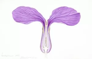
Background imageAnther Collection: Monkshood (Aconitum variegatum) C016 / 5690

Monkshood (Aconitum variegatum) C016 / 5690
Monkshood (Aconitum variegatum). Watercolour by Arthur Harry Church, 30 July 1906

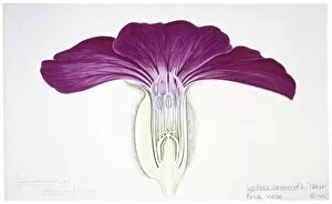
Background imageAnther Collection: Prick nose (Lychnis coronaria) C016 / 5687

Prick nose (Lychnis coronaria) C016 / 5687
Prick nose (Lychnis coronaria). Watercolour (Plate 171) by Arthur Harry Church, 12 July 1905

Background imageAnther Collection: Golden columbine (Aquilegia chrysantha) C016 / 5685

Golden columbine (Aquilegia chrysantha) C016 / 5685
Golden columbine (Aquilegia chrysantha). Ink and wash drawing by Arthur Harry Church, 1906. Figure for Types of floral mechanism. Natural History Museum, London, UK

Background imageAnther Collection: Honesty (Lunaria biennis), artwork C016 / 5676

Honesty (Lunaria biennis), artwork C016 / 5676
Hinesty (Lunaria biennis). Watercolour (Plate 146) by Arthur Harry Church. Held in the Botany Library at the Natural History Museum, London, UK

Background imageAnther Collection: Darlingtonia californica, artwork C016 / 5576

Darlingtonia californica, artwork C016 / 5576
California pitcherplant (Darlingtonia californica). Drawing (Plate 134.) by Arthur Harry Church, 1909. Held in the Botany Library at the Natural History Museum, London, UK

Background imageAnther Collection: Hardy larkspur (Delphinium formosum) C016 / 5575

Hardy larkspur (Delphinium formosum) C016 / 5575
Hardy larkspur (Delphinium formosum). Drawing by Arthur Harry Church, 1903. Held in the Botany Library at the Natural History Museum, London, UK

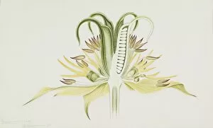
Background imageAnther Collection: Yellow fennel flower (Nigella orientalis) C016 / 5574

Yellow fennel flower (Nigella orientalis) C016 / 5574
Yellow fennel flower (Nigella orientalis). Drawing (Plate 062) by Arthur Harry Church, 1905. Held in the Botany Library at the Natural History Museum, London, UK

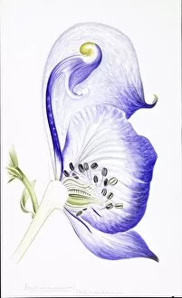
Background imageAnther Collection: Monk s-hood (Aconitumm napellus) C016 / 5572

Monk s-hood (Aconitumm napellus) C016 / 5572
Monk s-hood (Aconitumm napellus). Illustration (Plate 12) by Arthur Harry Church, 1903. Held in the Botany Library at the Natural History Museum, London, UK

Background imageAnther Collection: Centre of a hybrid tree peony flower C013 / 5129

Centre of a hybrid tree peony flower C013 / 5129
The centre of a flower of a hybrid tree peony. The genus Paeonia comprises both herbaceous and woody species (so-called tree peonies)

Background imageAnther Collection: Flower bud detail, SEM

Flower bud detail, SEM
Detail of inside a flower bud, coloured scanning electron micrograph (SEM). The flower is a cape fuchsia (Phygelius aequalis). Magnification is x14 at an image of 10cm wide

Background imageAnther Collection: Asiatic lily (Lilium sp. )

Asiatic lily (Lilium sp. )
Asiatic lily (Lilium sp.) flower


All Professionally Made to Order for Quick Shipping

"Exploring the Intricate World of Anthers: A Captivating Microscopic Journey" Delve into the mesmerizing realm of anthers, where nature's intricate beauty unfolds at a microscopic level. From the vibrant Flame lily (Gloriosa sp. ) to the delicate Geranium anther captured in stunning detail through scanning electron microscopy (SEM), this captivating journey takes us deep within the heart of flowers. Witness the Tea flower stamens as they reveal their enchanting structures under SEM, showcasing their unique patterns and textures. The Geranium pollen, magnified to perfection, showcases its fine intricacies that aid in pollination and reproduction. Marvel at the Forget-me-not flower's SEM image, unveiling its ethereal petals and stamen arrangement with unparalleled clarity. The Hong Kong orchid tree (Bauhinia blakeana) flowers from December bloom with grace and elegance, inviting us to appreciate their exquisite form. The Buttercup flower's SEM image reveals a world of miniature wonders hidden within its golden petals. Delicate Columbine flower stamens come alive under high-resolution SEM imaging, displaying their remarkable shapes and colors. A false-color SEM image captures the essence of a chickweed flower like never before—a visual feast for our senses. The majestic Amaryllis (Hippeastrum sp. ) stands tall with its resplendent blooms that captivate all who behold them. Finally, we encounter Thale cress flower through a micrograph—an intimate glimpse into nature's meticulous design process on a cellular scale. These diverse anthers remind us of Mother Nature's boundless creativity and her attention to even the tiniest details. Embark on this extraordinary expedition into anther exploration; let these images transport you into a world unseen by naked eyes but brimming with awe-inspiring beauty waiting to be discovered.