
Antigen Collection (page 2)
"Unveiling the Power of Antigens: Exploring the Dynamic Interplay with T Lymphocytes and Cancer Cells" In the realm of immunology


All Professionally Made to Order for Quick Shipping
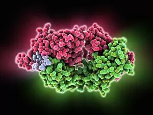
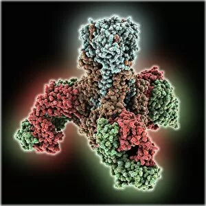
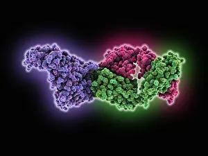
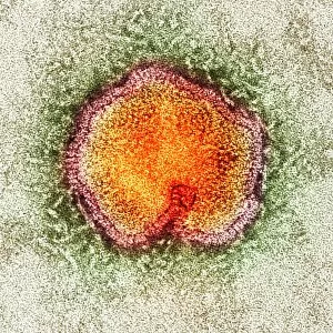
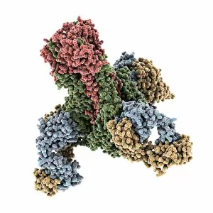
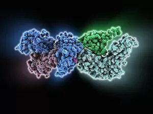
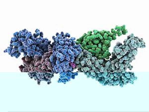
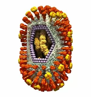
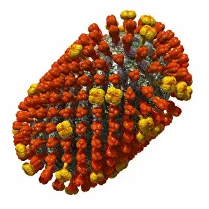
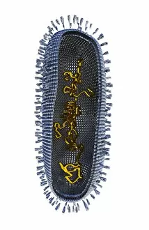
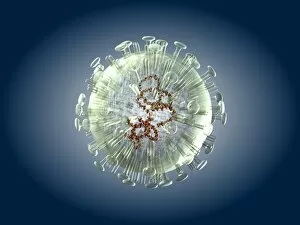
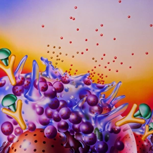
"Unveiling the Power of Antigens: Exploring the Dynamic Interplay with T Lymphocytes and Cancer Cells" In the realm of immunology, antigens hold a crucial role in our body's defense against various diseases. From Hepatitis B viruses to Hantavirus, these microscopic entities trigger an intricate immune response that safeguards our well-being. Through captivating SEM C001 / 1679 images, we witness T lymphocytes engaging in a fierce battle against cancer cells. These specialized white blood cells recognize specific antigens on malignant invaders, launching a targeted attack to eliminate them. Vaccination plays a pivotal role in preparing our immune system for future encounters. An awe-inspiring illustration showcases how vaccines stimulate our body's defenses by introducing microbes and their associated antigens. This prompts antibody production (such as Immunoglobulin G) and primes us for effective protection against potential infections. The foot-and-mouth disease virus (F006 / 9556) serves as another example where antigens play a central role. A conceptual image vividly portrays antibodies attaching themselves to bacteria, effectively neutralizing and killing them. Meanwhile, surrounding tissues become inflamed due to this battle, causing systemic effects that signal an ongoing fight within. Antihistamines come into play when allergies strike; they block histamine receptors from triggering allergic reactions caused by antigen exposure. This provides relief from symptoms like inflammation or itching – showcasing how understanding antigen interactions can lead to practical solutions for everyday health challenges. Nature's arsenal includes natural killer cells injecting toxins into bacteria—an extraordinary sight captured through scientific imaging techniques. These remarkable defenders contribute significantly to eliminating harmful pathogens from invading our bodies. Artistic representations beautifully depict the immune system's reaction when bacteria invade tissues—a synchronized dance between antibodies and foreign intruders unfolds before our eyes—showcasing nature's incredible ability to protect itself at its finest hour.