Fraud Blocker Skip to main content

Home > Science > SEM

Antigen presentation, SEM C016 / 3105

 : Antigen presentation, SEM C016  /  3105
In Stock
We accept all major payment options so you can pay for the things you need in the way that is most convenient for you

Choose Your Product

Framed Photos
Buy from AU$119.99
Photo Prints
Buy from AU$12.99
Jigsaw Puzzles
Buy from AU$89.99
Poster Prints
Buy from AU$24.99
Canvas Prints
Buy from AU$124.99
Fine Art Prints
Buy from AU$92.53
Metal Prints
Buy from AU$43.99
Cards
Buy from AU$12.99
Home Decor
Buy from AU$24.99

Science Photo Library Photo Prints and Wall Art

Antigen presentation, SEM C016 / 3105

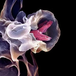
Antigen presentation. Coloured scanning electron micrograph (SEM) showing the interaction between a macrophage (blue) and a T helper lymphocyte (Th cell, yellow), two components of the bodys immune system. Both are types of white blood cell. Macrophages are antigen-presenting cells (APCs). They present antigens (fragments on the surface of pathogens or foreign objects) to T lymphocytes, activating them. Each T lymphocyte recognises and binds to a specific antigen. Binding of the Th cell to the antigen presented by the macrophage activates the Th cell. This leads to its proliferation and the activation of other immune cells that eliminate the antigen. Magnification: x7000 when printed 10cm wide. Science Photo Library features Science and Medical images including photos and illustrations. © STEVE GSCHMEISSNER/SCIENCE PHOTO LIBRARY

Media ID 9202945

Activating Activation Adaptive Immunity Antigen Colored Contact Defence Immune Immune Cell Immune System Immunity Immunological Immunology Interacting Interaction Leucocyte Leucocytes Leukocyte Leukocytes Macrophage Monocyte Presentation Presenting Specific Stimulating Stimulation System T Cell White Blood Cell Cells Cutouts Scanning Electron Microscope

FEATURES IN THESE COLLECTIONS

> Animals > Mammals > Muridae > Sua

> Science > SEM

SECURE PAYMENTS
We accept all major payment options so you can pay for the things you need in the way that is most convenient for you

Made in Australia
Safe Shipping, Easy Returns

FREE PERSONALISATION
We are proud to offer a range of customisation including Cropping, Captions, and Filters. Zoom Cropping can be adjusted in the basket


EDITORS COMMENTS

This print titled "Antigen Presentation" showcases the intricate interaction between a macrophage and a T helper lymphocyte, two vital components of the body's immune system. In this colored scanning electron micrograph (SEM), we witness the dynamic connection between these white blood cells that play crucial roles in defending our bodies against pathogens. The blue-hued macrophage, an antigen-presenting cell (APC), is responsible for presenting antigens—fragments found on the surface of foreign objects or pathogens—to T lymphocytes. The yellow-colored T helper lymphocyte, also known as Th cell, recognizes and binds to specific antigens presented by macrophages. This binding process activates the Th cell, leading to its proliferation and triggering other immune cells' activation to eliminate the antigen. With a magnification of x7000 when printed 10cm wide, this SEM image offers us a glimpse into the fascinating world of immunology and adaptive immunity. It highlights how different types of white blood cells work together harmoniously to protect our bodies from harmful invaders. Photographed by Steve Gschmeissner from Science Photo Library, this image beautifully captures both scientific precision and artistic appeal. It serves as a reminder of the complexity and resilience inherent in our immune system while offering insights into medical research and advancements aimed at understanding and harnessing its power for improved healthcare outcomes.


Framed Prints of Antigen presentation, SEM C016 / 3105

Discover the intricacies of the immune system with our captivating framed prints from Media Storehouse. Featuring the stunning image "Antigen Presentation, SEM C016 / 3105" by STEVE GSCHMEISSNER/SCIENCE PHOTO LIBRARY, this framed print showcases the interaction between a macrophage and a T helper lymphocyte in mesmerizing detail. The vibrant colors and high-resolution image provide an enlightening view into the complex process of antigen presentation. Bring this scientific masterpiece into your home or office to inspire curiosity and ignite conversations about the wonders of the human body.


Photo Prints of Antigen presentation, SEM C016 / 3105

Discover the intricacies of the immune system with our Media Storehouse Photographic Prints featuring the captivating image "Antigen presentation, SEM C016 / 3105" by STEVE GSCHMEISSNER/SCIENCE PHOTO LIBRARY. This coloured Scanning Electron Micrograph (SEM) showcases the interaction between a macrophage and a T helper lymphocyte, offering a unique and striking perspective on the antigen presentation process. Enhance your laboratory, classroom, or office space with this educational and visually stunning addition.


Poster Prints of Antigen presentation, SEM C016 / 3105

Discover the intricacies of the immune system with our captivating Media Storehouse Poster Prints. Featuring the striking image "Antigen presentation, SEM C016 / 3105" by STEVE GSCHMEISSNER/SCIENCE PHOTO LIBRARY, this poster offers a mesmerizing glimpse into the interaction between a macrophage and a T helper lymphocyte. This coloured scanning electron micrograph (SEM) illustrates the essential process of antigen presentation, showcasing the dynamic interplay between these immune cells. Bring the wonders of science into your workspace or learning environment with our high-quality, vibrant poster prints.


Jigsaw Puzzles of Antigen presentation, SEM C016 / 3105

Discover the intricacies of the immune system with our captivating jigsaw puzzles from Media Storehouse. This edition features the "Antigen Presentation" image (SEM C016 / 3105) by STEVE GSCHMEISSNER/SCIENCE PHOTO LIBRARY. Delve into the mesmerizing world of science as a macrophage (blue) and a T helper lymphocyte (Th cell, yellow) interact in this coloured scanning electron micrograph (SEM). Assemble the pieces to unravel the complex dance of the immune system, making learning an engaging and rewarding experience.

diamond
Made With High Grade Materials
thumb_up
Professional Quality Finish
package_2
Carefully Packed To Aid Safe Arrival

You May Also Like


Macrophage engulfing TB bacteria, SEMMacrophage engulfing TB bacteria, SEM
Add to Basket
Medical nanoparticles, conceptual image F005 / 0783Medical nanoparticles, conceptual image F005 / 0783
Add to Basket
Shigella bacterium and macrophage, SEM C016 / 8924Shigella bacterium and macrophage, SEM C016 / 8924
Add to Basket
Juvenile elephants (Loxodonta africana) at the David Sheldrick Elephant Orphanage, Nairobi National Park, Nairobi, Kenya, East Africa, AfricaJuvenile elephants (Loxodonta africana) at the David Sheldrick Elephant Orphanage, Nairobi National Park, Nairobi, Kenya, East Africa, Africa
Add to Basket
Print Shows A Member Of The Third EstatePrint Shows A Member Of The Third Estate
Add to Basket
Picture No. 10899380Picture No. 10899380
Add to Basket
Arsenal's Hector Bellerin and Emiliano Martinez Celebrate Community Shield Victory over ChelseaArsenal's Hector Bellerin and Emiliano Martinez Celebrate Community Shield Victory over Chelsea
Add to Basket
Ironwork DP131063Ironwork DP131063
Add to Basket
Chelsea Players Celebrate Second Goal Against Stoke City, Premier League 2016Chelsea Players Celebrate Second Goal Against Stoke City, Premier League 2016
Add to Basket
India / CalcuttaIndia / Calcutta
Add to Basket
Rangers Football Club 2003 Scottish Cup Champions Poppy Shirt: In Honor and RemembranceRangers Football Club 2003 Scottish Cup Champions Poppy Shirt: In Honor and Remembrance
Add to Basket
Anne Henriette of France (1727-1752), First half of the 19th centAnne Henriette of France (1727-1752), First half of the 19th cent
Add to Basket

Gallery Wall Inspiration

Skin section, SEM
SEM of section through human skin
Santos-Dumont / Sem
Neutrophil engulfing MRSA, SEM C018 / 8596
Candida fungus, SEM
Yeast fungus skin infection, SEM
Eyelash mites, SEM F006 / 9813
X and Y chromosomes
Lavender pollen grain, SEM
Scanning Electron Micrograph (SEM): Tardigrade or ‘Water Bear Magnification x 1250 (A4 size: 29. 7 cm width)
Nerve cell, SEM
Diatom, SEM
Cat tongue surface, SEM
False-coloured SEM (Scanning Electron Micrograph) of a Tardigrade (Tardigrada) amongst moss, from Peak District National Park, Derbyshire, UK. This tardigrade is approximately 0. 1 millimetres long. Digital composite
Collagen, Scanning electron micrograph (SEM)
T lymphocytes and cancer cell, SEM C001 / 1679
Tardigrade or Water Bear
Hazel pollen grains
Crysotile asbestos
Uterus lining during menstruation, SEM
Iris of the eye, SEM
Stylus in LP record
Salmonella bacteria, SEM
SEM of penicillin fungus
Vascular bundle, SEM
Geranium anther, SEM
Dahlia flower pollen, SEM


© Copyright Media Storehouse, All Rights Reserved 2003 - 2025
Facebook     Pinterest     100% Payment Secure     Reviews IO