
Leucocyte Collection
"Exploring the Mighty Leucocyte: Unveiling the Warriors of our Immune System" Leucocytes, also known as white blood cells


All Professionally Made to Order for Quick Shipping
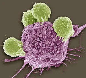
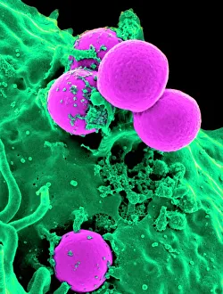
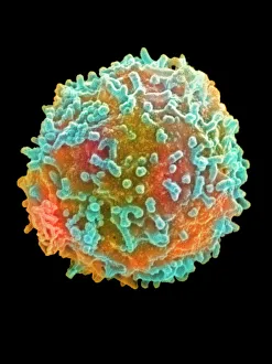
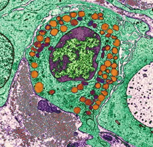
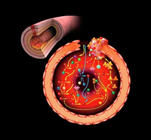
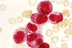
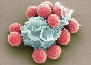
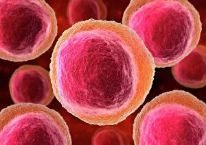
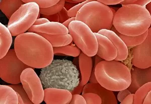
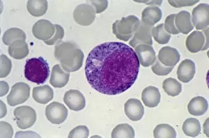
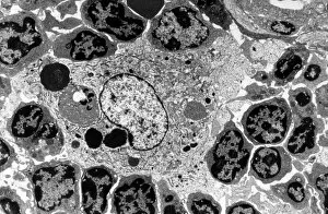
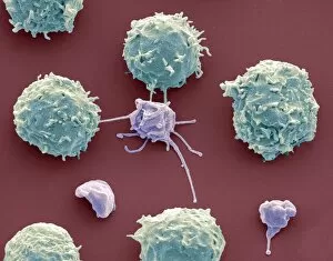
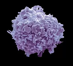
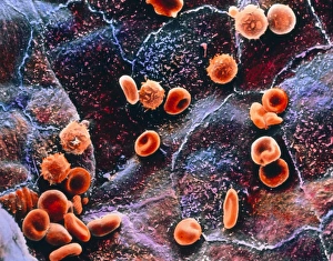
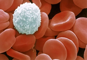
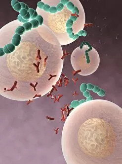
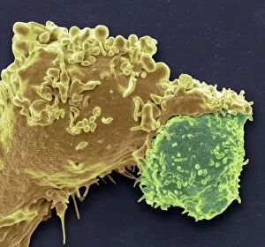
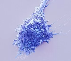
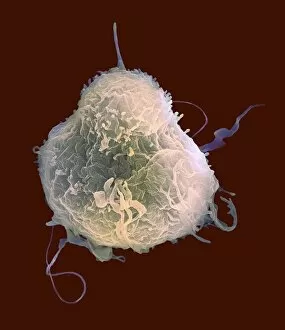
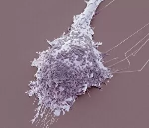
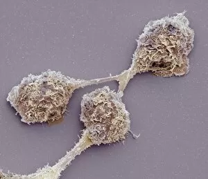
"Exploring the Mighty Leucocyte: Unveiling the Warriors of our Immune System" Leucocytes, also known as white blood cells, are the unsung heroes safeguarding our bodies from various threats. Among them, T lymphocytes play a crucial role in fighting cancer cells, acting as vigilant guardians against this formidable disease. (T lymphocytes and cancer cell, SEM C001 / 1679) Another remarkable the neutrophil - an expert at engulfing harmful bacteria like MRSA. Witness its extraordinary power under the microscope as it devours these dangerous microbes with precision and efficiency. (Neutrophil engulfing MRSA, SEM C018 / 8596) Intriguingly, human white blood cells bear HLA antigens that act as identification tags for immune recognition. Delve into their intricate structure through a detailed transmission electron microscopy image revealing their fascinating complexity. (TEM of human white blood cell bearing HLA antigen) Behold a vibrant world within our bloodstream. A colored scanning electron micrograph showcases a lymphocyte - one type of white blood cell - shining brightly amidst red and other cellular companions. (Colored SEM of a white blood cell [lymphocyte]) Basophilic leucocytes may be less well-known but possess significant functions in allergic reactions and inflammation responses within our bodies' defense mechanisms. Discover their unique characteristics through captivating microscopic imagery. (Basophil white blood cell) The intricate dance of life-saving clotting factors unfolds before us in an artwork depicting the mesmerizing beauty of the blood coagulation cascade—an essential process to prevent excessive bleeding when injuries occur. (Blood coagulation cascade, artwork C016 / 9873) Witness Dohle bodies within leucocytes—a microscopic phenomenon observed during certain infections or inflammatory conditions—captured vividly under high-resolution magnification.