Fraud Blocker Skip to main content

Home > Science > SEM

T lymphocytes and cancer cell, SEM C001 / 1679

T lymphocytes and cancer cell, SEM C001  /  1679
Sold 3
In Stock
We accept all major payment options so you can pay for the things you need in the way that is most convenient for you

Choose Your Product


Science Photo Library Photo Prints and Wall Art

T lymphocytes and cancer cell, SEM C001 / 1679

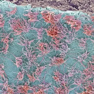
T lymphocytes and cancer cell. Coloured scanning electron micrograph (SEM) of T lymphocyte cells (green) attached to a cancer cell. T lymphocytes are a type of white blood cell that recognise a specific site (antigen) on the surface of cancer cells or pathogens and bind to it. Some T lymphocytes then signal for other immune system cells to eliminate the cell. The genetic changes that cause a cell to become cancerous lead to the presentation of tumour antigens on the cells surface. Magnification: x2300 when printed at 10 centimetres wide. Science Photo Library features Science and Medical images including photos and illustrations. © STEVE GSCHMEISSNER/SCIENCE PHOTO LIBRARY

Media ID 10842153

Antigen Antigenic Attached Attaching Attack Attacking Binding Bound Cancer Cancerous Cell Biology Cytology Cytotoxic Immune Response Immune System Immunity Immunology Leucocyte Leukocyte Lymphocytes Malignancy Malignant Oncology Recognising T Cell T Lymphocyte Tumour White Blood Cell Attach Bind Cells Recognise

SECURE PAYMENTS
We accept all major payment options so you can pay for the things you need in the way that is most convenient for you

Made in Australia
Safe Shipping, Easy Returns

FREE PERSONALISATION
We are proud to offer a range of customisation including Cropping, Captions, and Filters. Zoom Cropping can be adjusted in the basket


EDITORS COMMENTS

This photo print, titled "T lymphocytes and cancer cell" offers a mesmerizing glimpse into the intricate world of cellular interactions. In this colored scanning electron micrograph (SEM), we witness T lymphocyte cells (green) firmly attached to a cancer cell, showcasing the remarkable power of our immune system in combating malignancy. T lymphocytes, also known as T cells, are a vital component of our white blood cells. They possess the extraordinary ability to recognize specific antigens on the surface of cancerous cells or pathogens and bind to them. Once bound, these T lymphocytes signal other immune system cells to eliminate the targeted cell effectively. The genetic alterations that trigger cellular transformation into cancer result in tumor antigens being presented on their surfaces. This image magnified at x2300 when printed at 10 centimeters wide allows us to appreciate the minute details and complexity involved in this process. With its fusion of medicine, immunology, oncology, cytology, and cell biology disciplines captured through SEM technology by Steve Gschmeissner from Science Photo Library; this photograph serves as an invaluable resource for researchers and medical professionals alike. It sheds light on how our immune response works against malignant growths while providing insights into potential therapeutic approaches aimed at harnessing T lymphocyte's potent anti-cancer properties.


Framed Prints of T lymphocytes and cancer cell, SEM C001 / 1679

Introducing the Media Storehouse Framed Prints collection, featuring the captivating image "T lymphocytes and cancer cell, SEM C001 / 1679" by STEVE GSCHMEISSNER/SCIENCE PHOTO LIBRARY. This stunning, coloured scanning electron micrograph (SEM) showcases a T lymphocyte cell in vibrant green hues, engaged in an interaction with a cancer cell. This intriguing image is an excellent addition to any scientific or medical workspace, laboratory, or home decor that appreciates the beauty and complexity of the natural world. With our high-quality framed prints, you'll bring the wonders of science right into your living or working space.


Photo Prints of T lymphocytes and cancer cell, SEM C001 / 1679

Discover the intricacy of the immune system's defense against cancer with our exquisite Media Storehouse Photographic Print of "T Lymphocytes and Cancer Cell, SEM C001 / 1679" by Steve Gschmeissner/Science Photo Library. This coloured Scanning Electron Micrograph (SEM) captures the stunning detail of T lymphocyte cells (green) as they engage with a cancer cell, illustrating the complex interplay between the body's natural defense mechanisms and the development of malignant growths. Bring the wonders of scientific discovery into your home or office with this visually captivating and educational print.


Poster Prints of T lymphocytes and cancer cell, SEM C001 / 1679

Introducing the Media Storehouse range of stunning Poster Prints, featuring the captivating image "T Lymphocytes and Cancer Cell, SEM C001 / 1679" by STEVE GSCHMEISSNER/SCIENCE PHOTO LIBRARY from Science Photo Library. This mesmerizing scanning electron micrograph (SEM) showcases a unique interaction between T lymphocyte cells, represented in green, and a cancer cell. The intricate details and vibrant colors bring the complex world of immune system defense against cancer to life. Ideal for scientific research facilities, laboratories, classrooms, or any space that appreciates the beauty of scientific discovery, these high-quality poster prints are sure to inspire and educate.


Jigsaw Puzzles of T lymphocytes and cancer cell, SEM C001 / 1679

Discover the fascinating world of immune system defense with our captivating T Lymphocytes and Cancer Cell Jigsaw Puzzle from Media Storehouse. This intricately detailed puzzle, featuring an SEM image by STEVE GSCHMEISSNER/SCIENCE PHOTO LIBRARY, invites you to explore the complex relationship between T lymphocytes and cancer cells. Assemble the colorful pieces to reveal the stunning, high-definition image of green T lymphocyte cells engaged in their vital mission to combat cancer cells. A perfect addition to any home or classroom, this educational and visually stunning puzzle is sure to provide hours of engaging entertainment while expanding your knowledge of cellular biology.

diamond
Made With High Grade Materials
thumb_up
Professional Quality Finish
package_2
Carefully Packed To Aid Safe Arrival

You May Also Like


Brain surface, SEMBrain surface, SEM
Add to Basket
Tendon, SEMTendon, SEM
Add to Basket
Muscle fibre structure, artworkMuscle fibre structure, artwork
Add to Basket
King Penguins, South Georgia IslandKing Penguins, South Georgia Island
Add to Basket
Mongolia, KarakorumMongolia, Karakorum
Add to Basket
Anne Hathaways Cottage a98_05227Anne Hathaways Cottage a98_05227
Add to Basket
Ron Harris at Chelsea Soccer Training, Football League Division OneRon Harris at Chelsea Soccer Training, Football League Division One
Add to Basket
Prosthetic knees and obesity, X-ray C016 / 6596Prosthetic knees and obesity, X-ray C016 / 6596
Add to Basket
Muscles of the forearm, 1831 artworkMuscles of the forearm, 1831 artwork
Add to Basket
Trachea lining, SEM C015 / 9936Trachea lining, SEM C015 / 9936
Add to Basket
Snowflake, light micrographSnowflake, light micrograph
Add to Basket
VW Volkswagen Classic Camper van, 1965, Green, & whiteVW Volkswagen Classic Camper van, 1965, Green, & white
Add to Basket

Gallery Wall Inspiration

Nerve cell, SEM
False-coloured SEM (Scanning Electron Micrograph) of a Tardigrade (Tardigrada) amongst moss, from Peak District National Park, Derbyshire, UK. This tardigrade is approximately 0. 1 millimetres long. Digital composite
Santos-Dumont / Sem
Neutrophil engulfing MRSA, SEM C018 / 8596
X and Y chromosomes
Lavender pollen grain, SEM
Scanning Electron Micrograph (SEM): Tardigrade or ‘Water Bear Magnification x 1250 (A4 size: 29. 7 cm width)
Cat tongue surface, SEM
Hazel pollen grains
E. coli bacteria, SEM
Collagen, Scanning electron micrograph (SEM)
Scanning electron micrograph (SEM) of red blood cell
T lymphocytes and cancer cell, SEM C001 / 1679
Tardigrade or Water Bear
Crysotile asbestos
Uterus lining during menstruation, SEM
Iris of the eye, SEM
Stylus in LP record
Macrophage engulfing TB bacteria, SEM
Salmonella bacteria, SEM
SEM of penicillin fungus
Vascular bundle, SEM
Geranium anther, SEM
Dahlia flower pollen, SEM
Scanning Electron Micrograph (SEM): Praying Mantis - Magnification x 30 (if print A4 size: 29. 7 cm wide)
Tardigrade, SEM
Botanik Digitalis purpurea L. Fingerhut 160: 1


© Copyright Media Storehouse, All Rights Reserved 2003 - 2024
Facebook     Pinterest     100% Payment Secure     Reviews IO