
Apoptosis Collection
Apoptosis, the programmed cell death mechanism, is a captivating phenomenon that holds immense significance in various fields of research


All Professionally Made to Order for Quick Shipping
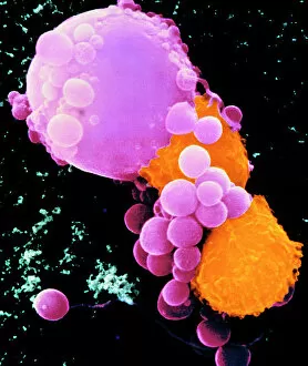
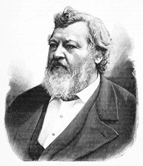
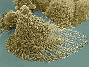
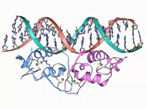
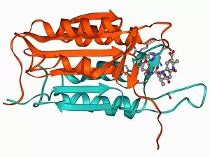
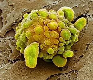
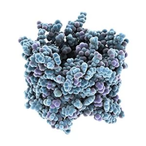
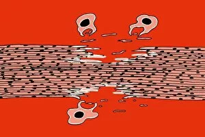
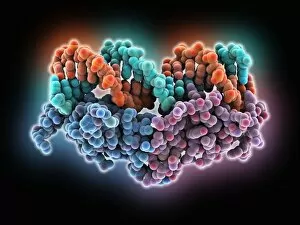
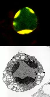
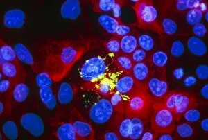
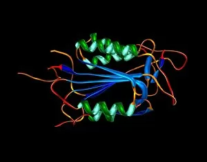
Apoptosis, the programmed cell death mechanism, is a captivating phenomenon that holds immense significance in various fields of research. This coloured scanning electron micrograph showcases lymphocytes launching an attack on a cancer cell, illustrating the intricate process at work. First discovered by Carl Vogt, a renowned German naturalist, it has since become an area of intense scientific exploration. The groundbreaking R&D 100 Award Winning Mitogenesis-Coupled Apoptosis Molecular Device (MCAMD) Team has made significant strides in unraveling the complexities surrounding this cellular event. Through their meticulous efforts and innovative approach, they have shed light on key players involved in apoptosis. The microscopic image of an apoptotic HeLa cell provides us with a glimpse into the structural changes occurring during this process. Additionally, we can observe Programmed Cell Death Protein molecules F006/9597 and F006/9548 as well as Transcription Factor and DNA molecule F006/9484 actively participating in orchestrating apoptosis. Caspase-3 and its inhibitor F006/9457 play crucial roles in regulating this intricate dance of life and death within cells. Similarly, Caspase-9 with its inhibitor depicted through molecular model F006/9442 adds another layer to our understanding of how these proteins function together harmoniously. Moreover, the presence of RuvBL1 helicase enzyme further emphasizes the complexity behind apoptosis regulation. Its involvement highlights yet another facet requiring extensive investigation to comprehend fully. Thanks to pioneers like Carl Vogt and cutting-edge researchers such as the MCAMD team who continue pushing boundaries within this field; we are gradually uncovering the mysteries surrounding apoptosis. These captivating images provide glimpses into a world where cells decide their own fate – whether to survive or undergo programmed self-destruction for the greater good.