Fraud Blocker Skip to main content

Asvd Collection

"Asvd: A Journey Through the Heart's Artistic Anatomy" Step into the captivating world of asvd

Background imageAsvd Collection: Heart bypass graft, artwork

Heart bypass graft, artwork
Heart bypass graft. Computer artwork of a heart that has had a blockage of the coronary arteries treated by coronary artery bypass graft (CABG) surgery

Background imageAsvd Collection: Heart bypass graft, artwork

Heart bypass graft, artwork
Heart bypass graft. Computer artwork of a heart that has had a blockage of the coronary arteries treated by coronary artery bypass graft (CABG) surgery

Background imageAsvd Collection: Blocked artery, computer artwork

Blocked artery, computer artwork
Blocked artery. Computer artwork of a clot of blood cells (thrombus, red) in an artery affected by atherosclerosis. This is a narrowing of an artery due to fatty deposits (yellow) on its inner walls

Background imageAsvd Collection: Aortic dissection, 3D CT scan

Aortic dissection, 3D CT scan
Aortic dissection. 3D computed tomography (CT) scan of a 67-year-old man with an aortic dissection. The aorta, the bodys main artery, is running vertically down centre (red)

Background imageAsvd Collection: Atherosclerosis, 3D CT scan

Atherosclerosis, 3D CT scan
Atherosclerosis. 3D computed tomography (CT) angiogram of the main blood vessels of the lower body a 58-year-old man, showing numerous atheromatous plaques (light, centre) on the vessel walls

Background imageAsvd Collection: Atherosclerosis, 3D CT scan C016 / 6537

Atherosclerosis, 3D CT scan C016 / 6537
Atherosclerosis. Coloured 3D computed tomography (CT) angiogram of the hip of a 67 year old patient with hypertension (high blood pressure) and hypercholesterolemia (high blood cholesterol)

Background imageAsvd Collection: Atherosclerosis, 3D CT scan C016 / 6540

Atherosclerosis, 3D CT scan C016 / 6540
Atherosclerosis. Coloured 3D computed tomography (CT) angiogram of the abdomen of a 73 year old patient. The aorta (cream, down centre), the bodys main artery, has calcified fatty plaques (atheroma)

Background imageAsvd Collection: Atherosclerosis in artery, artwork C016 / 6571

Atherosclerosis in artery, artwork C016 / 6571
Atherosclerosis in artery. Artwork of a longitudinal section through an artery that has been narrowed by atherosclerosis. Narrowing is referred to as stenosis

Background imageAsvd Collection: Atherosclerosis, 3D CT scan C016 / 6538

Atherosclerosis, 3D CT scan C016 / 6538
Atherosclerosis. Coloured 3D computed tomography (CT) angiogram of the hip of a 67 year old patient with hypertension (high blood pressure) and hypercholesterolemia (high blood cholesterol)

Background imageAsvd Collection: Atherosclerosis, 3D CT scan C016 / 6539

Atherosclerosis, 3D CT scan C016 / 6539
Atherosclerosis. Coloured 3D computed tomography (CT) angiogram of the heart of a 69 year old patient that has had an aortic heart valve replacement

Background imageAsvd Collection: Narrowed artery, artwork

Narrowed artery, artwork
Narrowed artery. Computer artwork of a section through an artery (horizontal, centre) affected by atherosclerosis, showing red blood cells (red) and fat deposits (cream)

Background imageAsvd Collection: Atherosclerosis, X-ray

Atherosclerosis, X-ray
Atherosclerosis. X-ray of a patient with atherosclerosis of the abdominal aorta (white, down centre). The aorta is the bodys main artery, it carries blood from the heart to the rest of the body

Background imageAsvd Collection: Atheromatous artery, artwork C015 / 6445

Atheromatous artery, artwork C015 / 6445
Atheromatous artery. Computer artwork showing the interior of an artery affected by atherosclerosis. Atherosclerosis is narrowing of an artery (stenosis) due to fatty deposits (beige)

Background imageAsvd Collection: Atheromatous artery, artwork C015 / 6446

Atheromatous artery, artwork C015 / 6446
Atheromatous artery. Cutaway computer artwork showing the interior of an artery affected by atherosclerosis. Atherosclerosis is narrowing of an artery (stenosis) due to fatty deposits (beige)

Background imageAsvd Collection: Atheromatous artery C013 / 4612

Atheromatous artery C013 / 4612
Atheromatous artery. Cutaway computer artwork showing the interior of an artery affected by atherosclerosis, showing red blood cells (red)

Background imageAsvd Collection: Doppler ultrasound scanning of carotid artery

Doppler ultrasound scanning of carotid artery

Background imageAsvd Collection: Narrowed coronary artery

Narrowed coronary artery

Background imageAsvd Collection: Angina pectoris

Angina pectoris. Illustration of a heart diseased with the cardiac condition angina pectoris. The heart is outlined in blue, with the aortic arch at top (red) and coronary arteries at lower frame

Background imageAsvd Collection: Colour SEM of atherosclerosis in coronary artery

Colour SEM of atherosclerosis in coronary artery
Atherosclerosis. Coloured Scanning Electron Micro- graph (SEM) of a longitudinal section through a human coronary artery of the heart showing athero- sclerosis

Background imageAsvd Collection: Artwork showing atherosclerosis of coronary artery

Artwork showing atherosclerosis of coronary artery
Coronary artery disease. Illustration of a human heart showing narrowing of the coronary artery due to atherosclerosis - build up of fatty deposits (atheroma, yellow)

Background imageAsvd Collection: Heart disease: hypertension, atheroma, lung oedema

Heart disease: hypertension, atheroma, lung oedema

Background imageAsvd Collection: Artwork of heart attack due to atherosclerosis

Artwork of heart attack due to atherosclerosis
Heart attack. Artwork of a human heart during a heart attack (myocardial infarction). Here, an embolus (red, in the sectioned artery at lower right)

Background imageAsvd Collection: Heart bypass grafts

Heart bypass grafts. Artwork of a heart that has had a blockage of the coronary arteries treated by coronary artery bypass graft (CABG) surgery

Background imageAsvd Collection: Narrowed artery due to cholesterol

Narrowed artery due to cholesterol

Background imageAsvd Collection: Atherosclerosis

Atherosclerosis. Artwork of a human heart showing narrowing of the coronary arteries due to atherosclerosis, a build up of fatty deposits (atheroma, yellow) on the artery wall

Background imageAsvd Collection: Narrowed artery, computer artwork

Narrowed artery, computer artwork
Narrowed artery. Computer artwork of a narrowed artery, showing red blood cells (red) and high levels of low density lipoprotein (LDL, yellow balls)

Background imageAsvd Collection: Blocked artery

Blocked artery
Artery disease. Computer artwork of blood cells (round) in an artery affected by atherosclerosis. This is a narrowing of an artery due to fatty deposits (yellow) on its inner walls

Background imageAsvd Collection: Arterial bypass

Arterial bypass. Artwork of a bypass around a narrowed region of the right femoral artery (red, down centre) of the human thigh

Background imageAsvd Collection: Artwork of artery narrowed by atherosclerosis

Artwork of artery narrowed by atherosclerosis
Atherosclerosis. Illustration of a cross-section through an artery narrowed by atherosclerosis. Fat build-up (yellow) has narrowed the lumen (channel) of the artery thus restricting the blood flow

Background imageAsvd Collection: Artwork of narrowed artery by atherosclerosis

Artwork of narrowed artery by atherosclerosis
Atherosclerosis. Artwork illustrating a cross section through an artery narrowed by atherosclerosis. A raised yellow plaque is seen in the wall of the artery

Background imageAsvd Collection: Illustration of narrowed artery in hyperlipidaemia

Illustration of narrowed artery in hyperlipidaemia
Hyperlipidaemia. Illustration of the narrowing of an artery (atherosclerosis) due to high levels of lipids in the blood (hyperlipidaemia)

Background imageAsvd Collection: High blood cholesterol

High blood cholesterol. Artwork of hypercholester- olaemia or hyperlipidaemia, high levels of the lipid cholesterol in the blood

Background imageAsvd Collection: LM of cross-section of artery with atherosclerosis

LM of cross-section of artery with atherosclerosis
Atherosclerosis. Light micrograph of a cross section through an artery obstructed with atheroma plaque. The disease is known as atherosclerosis

Background imageAsvd Collection: Aorta affected by atherosclerosis & normal one

Aorta affected by atherosclerosis & normal one
Atherosclerosis. Cross-sections showing a comparison between a healthy aorta (bottom), the main artery of the body, and one showing several plaques (yellow-white) of atheroma

Background imageAsvd Collection: Blocked coronary artery, artwork

Blocked coronary artery, artwork
Blocked coronary artery. Computer artwork of a clot (red) in a coronary artery. The clot has been caused by atherosclerosis, the build up of fatty plaques (yellow) on the artery wall

Background imageAsvd Collection: Heart ischaemia

Heart ischaemia
Heart disease. Artwork showing a heart with ischaemia (oxygen shortage) due to a blocked artery. The cardiac tissue beneath the blockage (grey)

Background imageAsvd Collection: Heart disease caused by fatty foods

Heart disease caused by fatty foods
Heart disease caused by unhealthy food. Conceptual artwork of unhealthy foods being forced into blood vessels near the heart

Background imageAsvd Collection: Coronary artery disease

Coronary artery disease. Computer artwork of red blood cells flowing through an increasingly (top to bottom) blocked coronary artery

Background imageAsvd Collection: Narrowed abdominal arteries, angiogram

Narrowed abdominal arteries, angiogram
Narrowed abdominal arteries, coloured angiogram (blood vessel X-ray). The aorta, the bodys main artery, is at top. It branches into two iliac arteries (bottom)

Background imageAsvd Collection: High cholesterol levels

High cholesterol levels. Computer artwork of balls of cholesterol. When there are high levels of low density lipoprotein (LDL)

Background imageAsvd Collection: Atherosclerosis in femoral arteries X-ray

Atherosclerosis in femoral arteries X-ray
Coloured X-ray (front view) of the pelvic region in a 72 year old patient showing atherosclerosis (mottled appearance) in the femoral arteries running on the inside of the femur (thigh) bones

Background imageAsvd Collection: Vascular disease progression, artwork

Vascular disease progression, artwork
Vascular disease progression. Artwork showing the progression of vascular disease in cross-section (left) and transverse section (right). At top is a healthy artery

Background imageAsvd Collection: Abdominal aortic aneurysm, 3D CT scan

Abdominal aortic aneurysm, 3D CT scan

Background imageAsvd Collection: Stenosis of carotid artery, CT scan

Stenosis of carotid artery, CT scan
Stenosis of carotid artery. Coloured three-dimensional computed tomography (CT) angiography scan of the neck arteries (red) of a 41-year-old patient, showing a stenosis (narrowing)

Background imageAsvd Collection: Nanobots and atherosclerosis, artwork

Nanobots and atherosclerosis, artwork
Nanobots and atherosclerosis. Artwork of nanorobots inside a blood vessel as they carry out treatment of atherosclerosis. Blood vessels can become blocked or narrowed by the build-up of fatty plaques

Background imageAsvd Collection: Iliac artery stenosis, 3-D MRI scan

Iliac artery stenosis, 3-D MRI scan
3-D Magnetic resonance imaging (MRI) scan of the abdomen of a patient, showing stenosis (narrowing, centre right) in one of the two iliac arteries which lead into the legs from the abdominal aorta

Background imageAsvd Collection: Bilateral renal artery stenosis, MRA sca

Bilateral renal artery stenosis, MRA sca
Coloured three-dimensional Magnetic Resonance Angiogram (MRA) scan of the abdomen of a male patient aged 77, showing bilateral renal artery stenosis (narrowing)

Background imageAsvd Collection: Cholesterol, molecular model

Cholesterol, molecular model
Cholesterol. Molecular model of the fatty and waxy alcohol cholesterol. Atoms are represented by spheres and are colour-coded: carbon (black), hydrogen (grey), oxygen (red)


All Professionally Made to Order for Quick Shipping

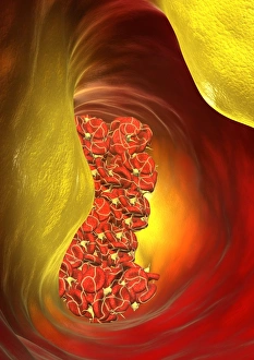
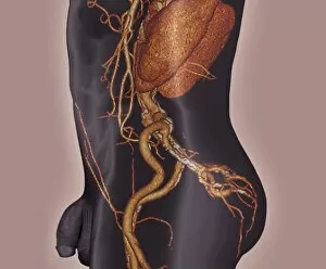
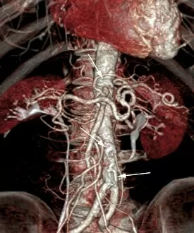
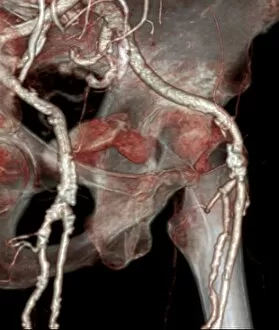
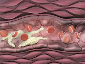
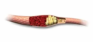
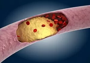
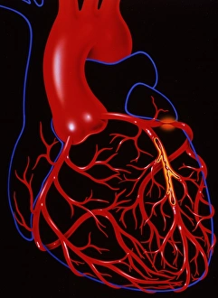
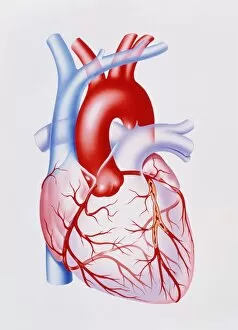
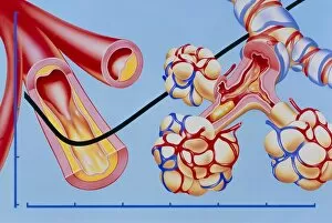
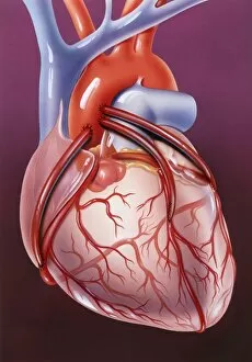
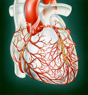
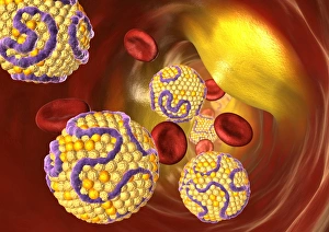
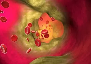
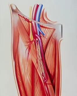
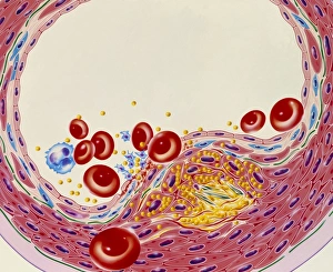
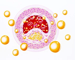
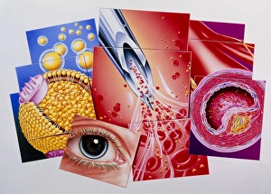
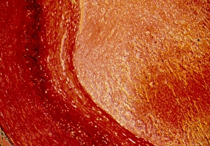
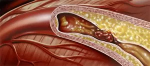
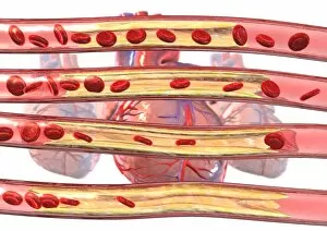
"Asvd: A Journey Through the Heart's Artistic Anatomy" Step into the captivating world of asvd, where medical science and artistic expression intertwine to showcase the intricate beauty of our cardiovascular system. From heart bypass grafts to blocked arteries, this collection of artwork and computer-generated imagery takes us on a visual voyage through various cardiac conditions. In one frame, we witness the meticulous craftsmanship of a heart bypass graft, a surgical procedure that redirects blood flow around a blocked artery. The delicate precision required for such an operation is mirrored in the artist's brushstrokes, capturing both the technicality and artistry involved in saving lives. Moving onto computer artwork, we encounter an awe-inspiring depiction of a blocked artery. With its vibrant colors contrasting against dark shadows, it serves as a reminder of how even within our bodies' most vital pathways can lie hidden dangers that require immediate attention. Aortic dissection comes alive through a mesmerizing 3D CT scan. This immersive experience allows us to explore every nook and cranny of this life-threatening condition with unparalleled clarity. It unveils not only the severity but also highlights advancements in medical imaging technology that aid doctors in diagnosing and treating such complex ailments. The journey continues with another 3D CT scan showcasing atherosclerosis – plaque buildup within arteries – which remains one of society's leading health concerns. These scans provide invaluable insights into disease progression while simultaneously evoking wonder at how something so microscopic can have such profound consequences on our overall well-being. Artwork further captures the essence of atherosclerosis by depicting its presence within an artery; here again, we see creativity merging seamlessly with scientific accuracy to portray this silent yet formidable adversary lurking beneath our skin. X-ray images shed light on yet another aspect of asvd—narrowed arteries—a sight often associated with restricted blood flow and potential complications like heart attacks or strokes. These stark visuals serve as reminders for early detection and proactive measures to maintain a healthy cardiovascular system.